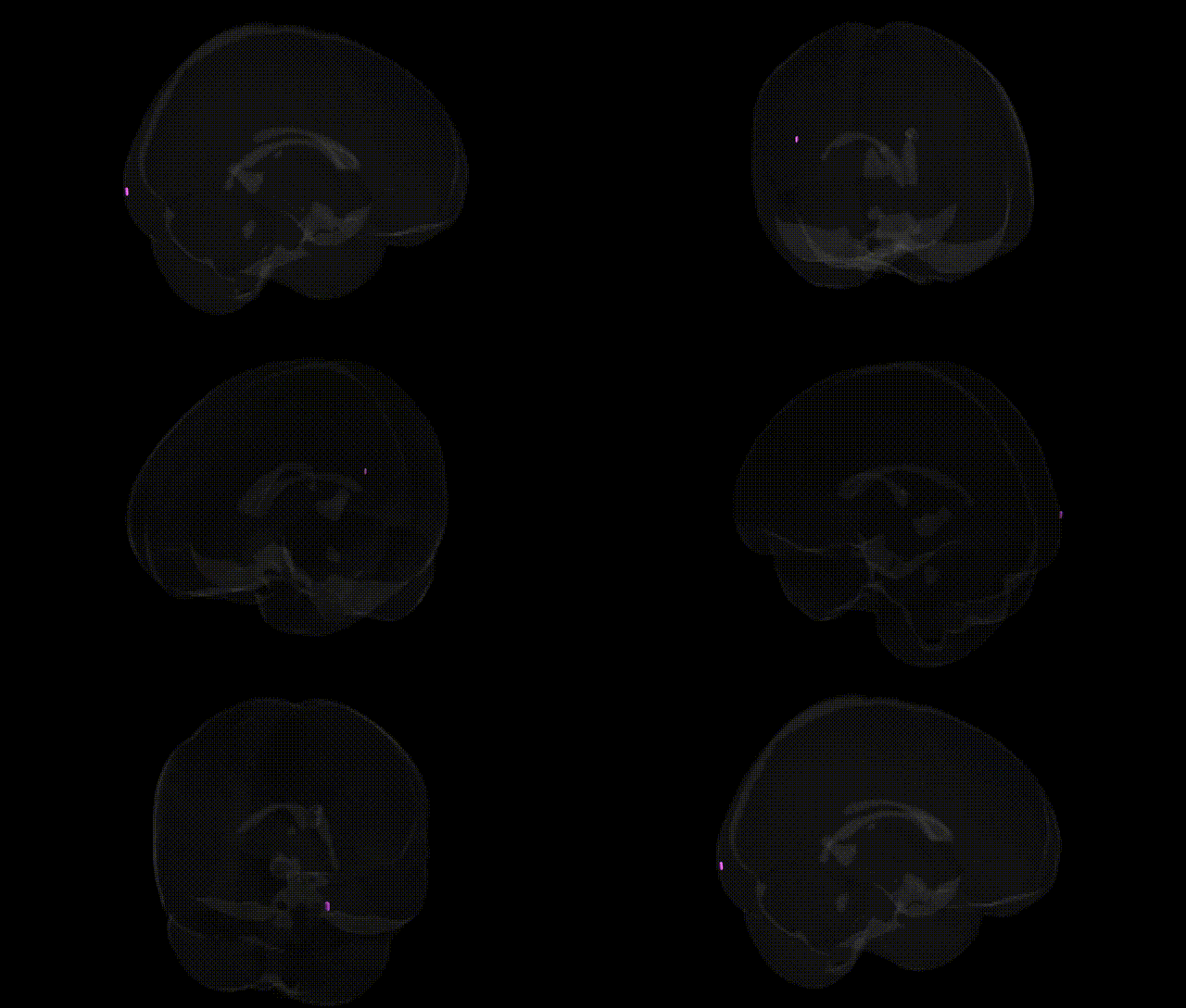
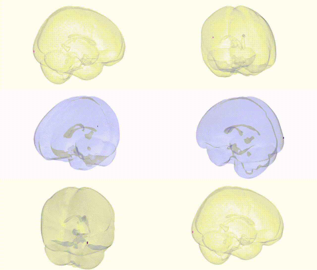
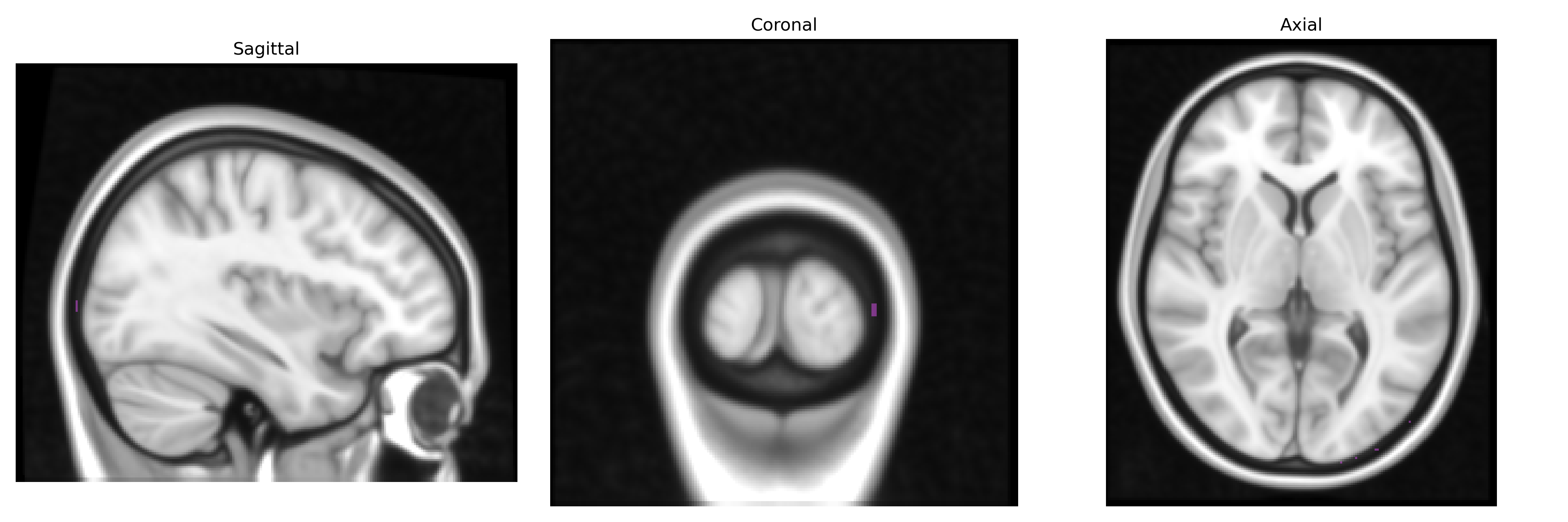
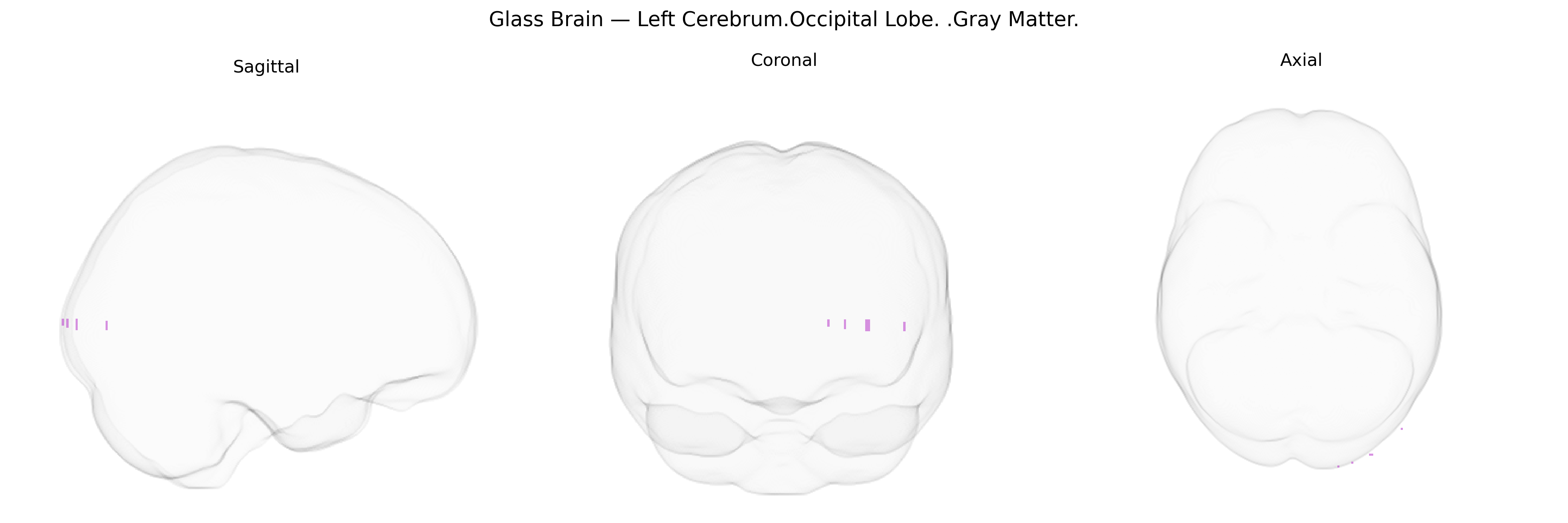
The bilateral Left Cerebrum Occipital Lobe Gray Matter corresponds to cortical regions within the left occipital lobe that primarily subserve visual processing, including basic pattern, color, motion, and spatial information analysis. This territory encompasses primary visual cortex (striate cortex) as well as surrounding extrastriate areas that contribute to higher-order visual perception and integration with dorsal and ventral visual streams. Neuronal populations here receive thalamic input from the lateral geniculate nucleus and project to associative visual and multimodal cortical regions, supporting processes such as visual recognition, visuospatial orientation, and visually guided behavior. Lesions in this gray matter region can lead to contralateral visual field deficits, visual agnosias, or disturbances in visual perception depending on the specific subregions affected. Occipital lobe
The bilateral left occipital lobe gray matter (as labeled in the Talairach 1 mm atlas) is a core visual processing region whose structure and function show substantial heritability, with twin and family studies indicating strong genetic contributions to occipital cortical thickness, surface area, and volume; GWAS have implicated loci in genes involved in neurodevelopment, synaptic function, and myelination (for example variants near or within PAX6, EPHB, FGFR, and cell-adhesion genes) in shaping occipital morphology and visual cortex metrics. Large-scale imaging–genetics consortia such as ENIGMA and UK Biobank have identified common variants associated with occipital lobe gray matter measures that overlap with risk loci for neurodevelopmental and psychiatric conditions, including schizophrenia, autism spectrum disorders, ADHD, and major depression, where altered occipital thickness or volume has been observed as an intermediate phenotype. Occipital gray matter variation has also been linked genetically to visual acuity and refractive errors, migraine (particularly visual aura), and traits involving visual processing or reading, with some studies suggesting that alleles affecting occipital structure and connectivity contribute to susceptibility to cortical visual disorders, hallucinations, and photosensitive epilepsy. Across these findings, the left occipital cortex frequently appears in polygenic architectures rather than through single strong-effect loci, acting as a genetically influenced substrate that mediates the impact of widespread neurodevelopmental and synaptic genes on visual and higher-order cognitive phenotypes.
Overview generated by GPT-4o (2026).
Region ID: 519
Hemisphere: bilateral
Atlas: Talairach labels 1mm
Full Quality Version: Download MP4
Full Quality Version: Download MP4
Wali Sidiqyar*, Gaurav Rudravaram*, Elyssa M. McMaster, Trent M. Schwartz, Adam M. Saunders, Kurt G. Schilling, Bennett A. Landman "Introducing SPINS: A Shared Public Visualization Library of Neuroanatomical Structures." Medical Imaging with Deep Learning- short paper
This resource is licensed under CC0 1.0 Universal (Public Domain).