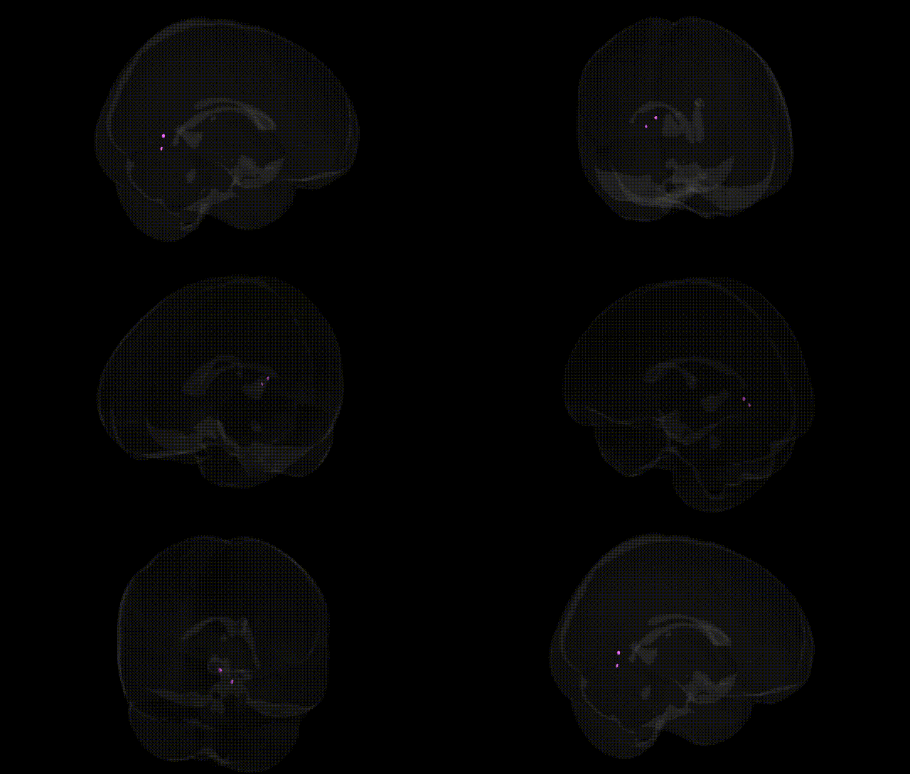
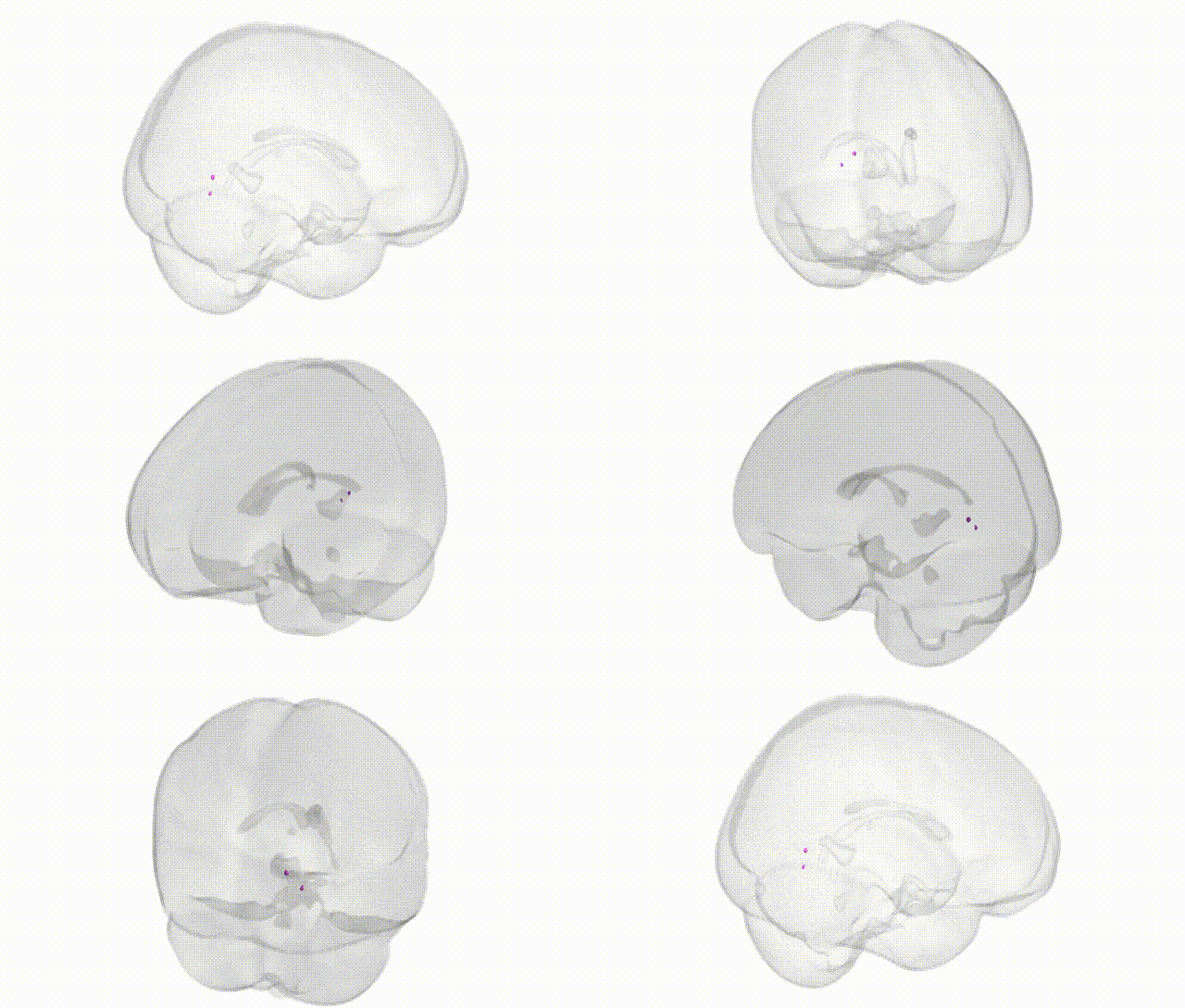
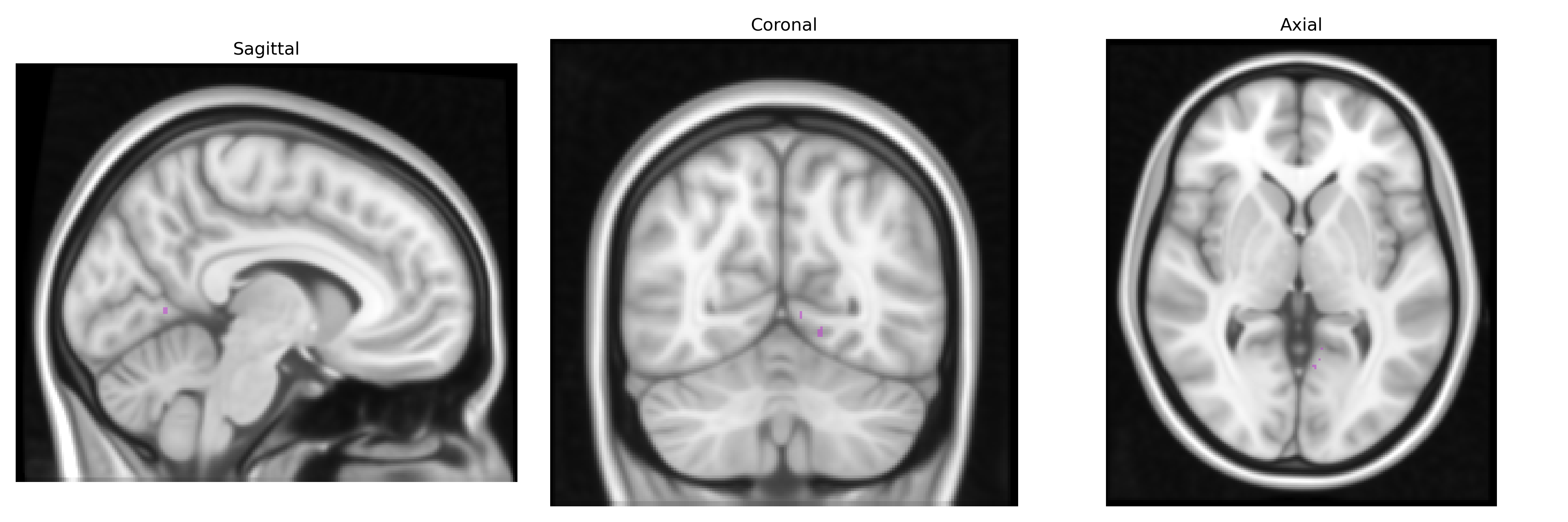
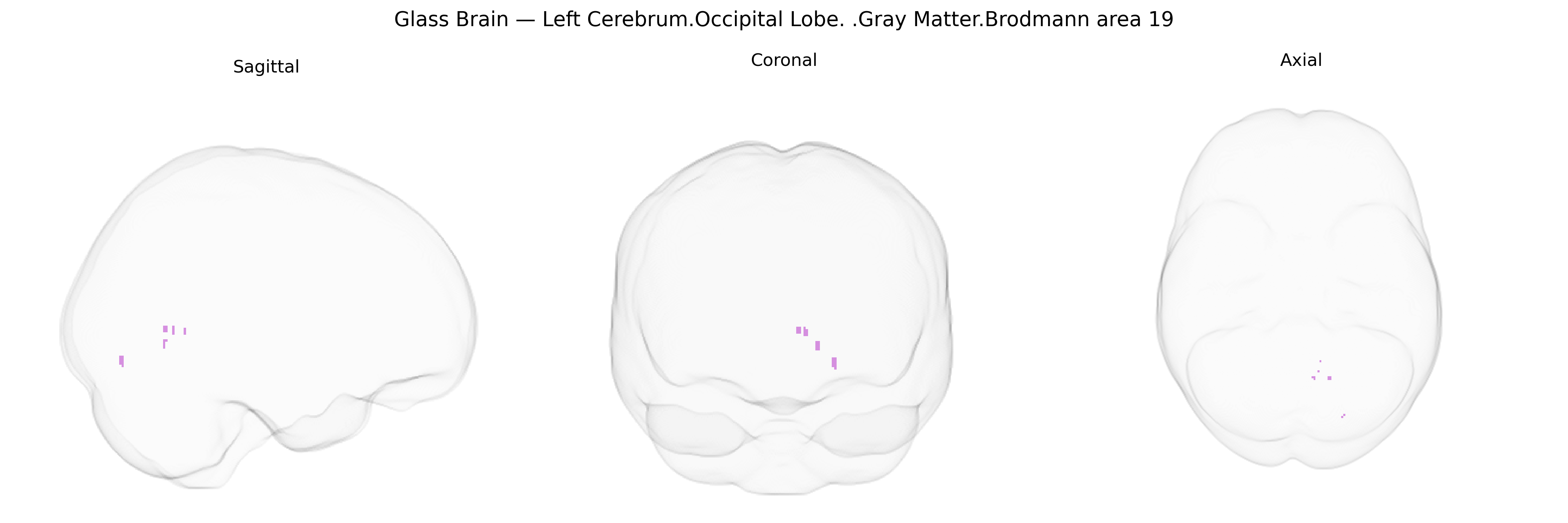
The bilateral Left Cerebrum Occipital Lobe Gray Matter Brodmann area 19 corresponds to higher-order visual association cortex located in the lateral and medial portions of the occipital lobe, adjacent to primary (Brodmann area 17) and secondary (Brodmann area 18) visual areas. This region integrates complex visual information, including form, color, motion, and object features, and contributes to visual recognition, spatial perception, and the interpretation of visual scenes. Neuronal populations here receive convergent input from earlier visual areas and relay processed information to temporal and parietal cortices, supporting functions such as object identification and visuospatial analysis. Its cytoarchitecture is characterized by a well-developed granular layer and multiple distinct pyramidal layers typical of association cortex. There is no direct link for Brodmann area 19; see the related region Visual cortex.
Brodmann area 19 (associative visual cortex in the occipital lobe) has been implicated in several genetic and GWAS-based findings, largely through imaging-genetics and disorder-trait association studies. Structural and functional variation in this region has been linked to polymorphisms in genes involved in neurodevelopment, synaptic plasticity, and myelination, including BDNF (e.g., Val66Met), NRG1, and genes regulating axon guidance and visual pathway development; large-scale MRI GWAS (e.g., ENIGMA and UK Biobank) have identified common variants influencing occipital cortical thickness and surface area in loci such as HMGA2 and other brain-structure–associated genes. BA19 shows altered gray matter or activation patterns in genetically influenced disorders like schizophrenia, autism spectrum disorder, and major depression, with risk variants in genes such as COMT, DISC1, and multiple GWAS-identified schizophrenia loci contributing to visual processing and higher-order perceptual abnormalities. Genetic factors contributing to migraine with aura, occipital lobe epilepsy, and visual hallucinations (including in Parkinson’s disease and Lewy body dementia) have also been associated with altered structure or function in the occipital association cortex, although specific causal variants often act via broader cortical or network-level mechanisms rather than BA19 alone.
Overview generated by GPT-4o (2026).
Region ID: 264
Hemisphere: bilateral
Atlas: Talairach labels 1mm
Full Quality Version: Download MP4
Full Quality Version: Download MP4
Wali Sidiqyar*, Gaurav Rudravaram*, Elyssa M. McMaster, Trent M. Schwartz, Adam M. Saunders, Kurt G. Schilling, Bennett A. Landman "Introducing SPINS: A Shared Public Visualization Library of Neuroanatomical Structures." Medical Imaging with Deep Learning- short paper
This resource is licensed under CC0 1.0 Universal (Public Domain).