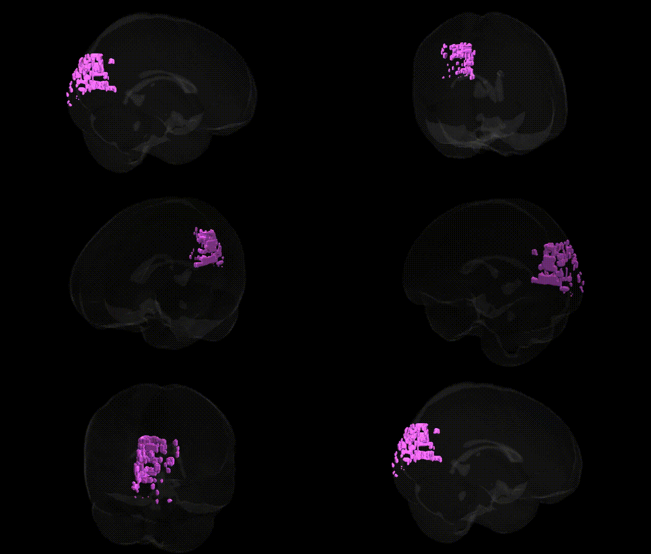
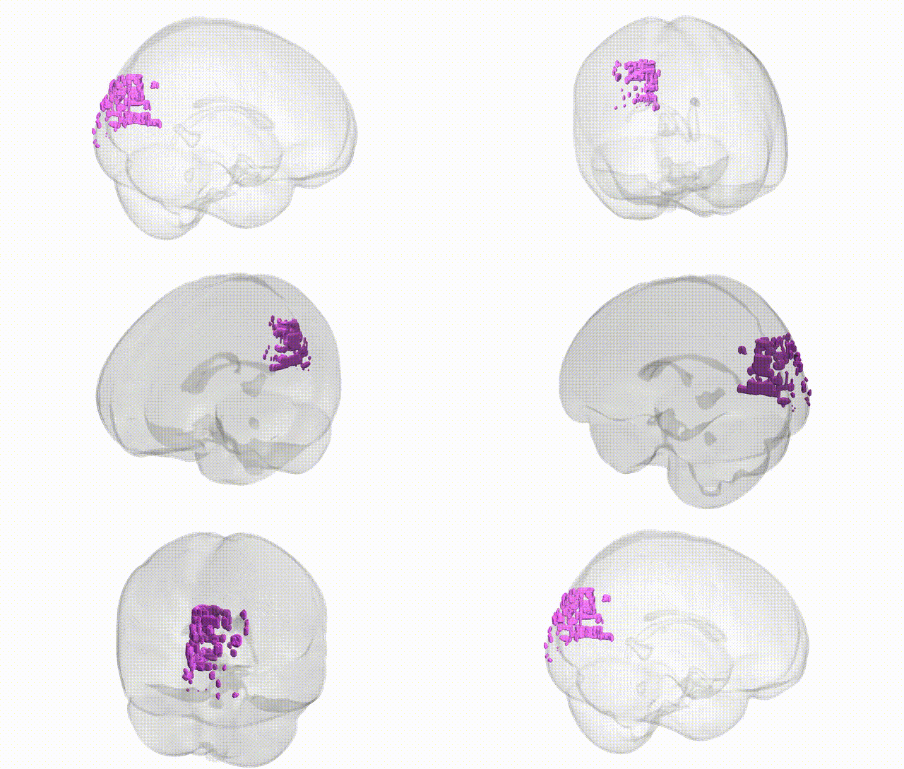
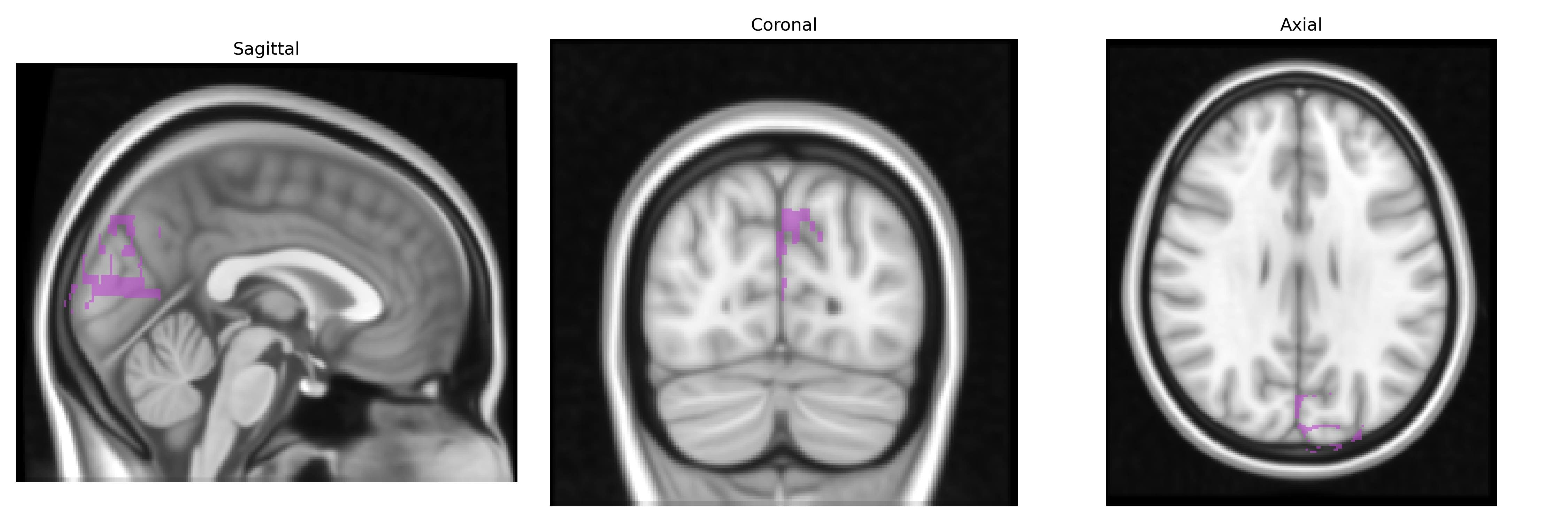
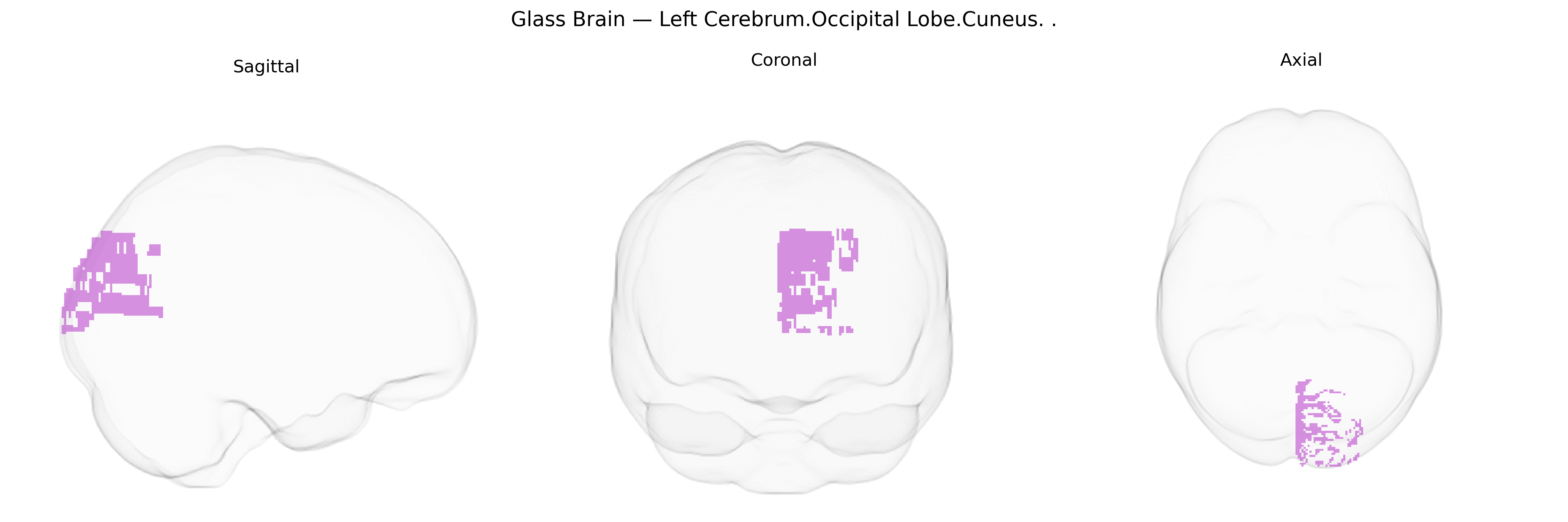
The bilateral Left Cerebrum.Occipital Lobe.Cuneus corresponds to the cuneus, a wedge-shaped cortical region of the medial occipital lobe located between the parieto‑occipital sulcus and the calcarine sulcus. It forms part of the primary and associative visual cortices, receiving input via the geniculostriate pathway and contributing to basic visual processing such as pattern, motion, and spatial feature analysis, particularly from the lower visual field. The cuneus participates in early visual integration, interactions with higher-order parietal and temporal regions for visuospatial perception and attention, and has been implicated in visual imagery and certain aspects of visual memory.
The bilateral left cerebrum occipital lobe cuneus, a core visual-processing region, has not itself been the explicit focus of many region-specific GWAS, but convergent imaging-genetics and large-scale GWAS/meta-analyses implicate it in several heritable traits and disorders. Cortical thickness and surface area in the cuneus show substantial SNP-based heritability and are influenced by common variants in genes involved in neurodevelopment and synaptic function (e.g., loci near HMGA2, KIAA0586, and regulatory regions for Wnt and axon-guidance pathways), as reported in ENIGMA and UK Biobank imaging-genetics studies. Genetic liability for major depressive disorder and schizophrenia has been associated with structural and functional alterations in the cuneus, with polygenic risk scores for these disorders correlating with reduced occipital/cuneus volume or altered activation during visual and cognitive tasks. Variants linked to migraine (such as those near TRPM8 and genes in glutamatergic and vascular pathways) have been related to occipital cortex and cuneus functional changes, consistent with visual aura and photophobia phenotypes. In addition, genetic influences on general cognitive ability, reading and visual perception, and neuroticism or anxiety have been associated with cuneus morphology or activation patterns in imaging-genetics work, suggesting that polygenic architectures for these traits partly act through development and functional modulation of this occipital lobe region.
Overview generated by GPT-4o (2026).
Region ID: 473
Hemisphere: bilateral
Atlas: Talairach labels 1mm
Full Quality Version: Download MP4
Full Quality Version: Download MP4
Wali Sidiqyar*, Gaurav Rudravaram*, Elyssa M. McMaster, Trent M. Schwartz, Adam M. Saunders, Kurt G. Schilling, Bennett A. Landman "Introducing SPINS: A Shared Public Visualization Library of Neuroanatomical Structures." Medical Imaging with Deep Learning- short paper
This resource is licensed under CC0 1.0 Universal (Public Domain).