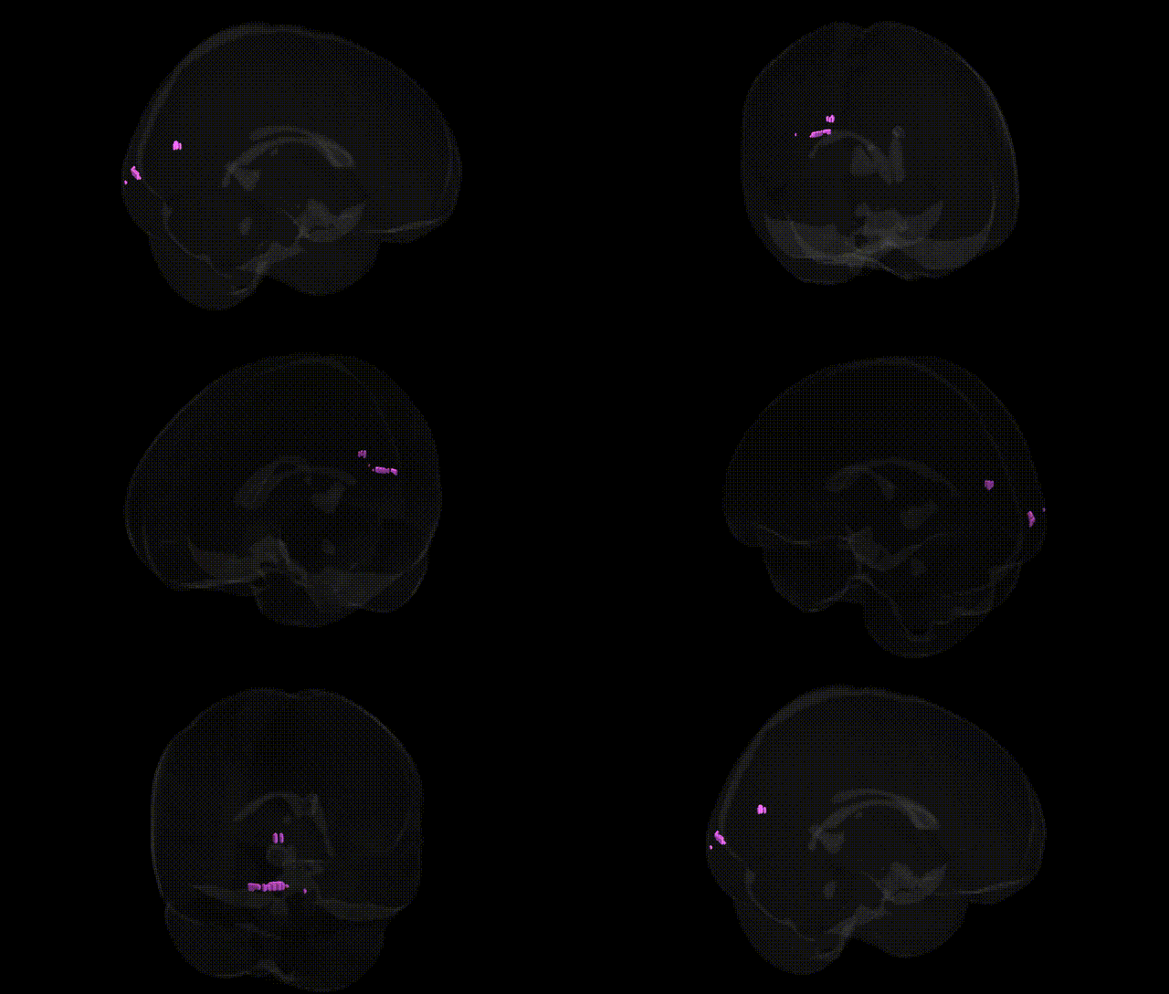
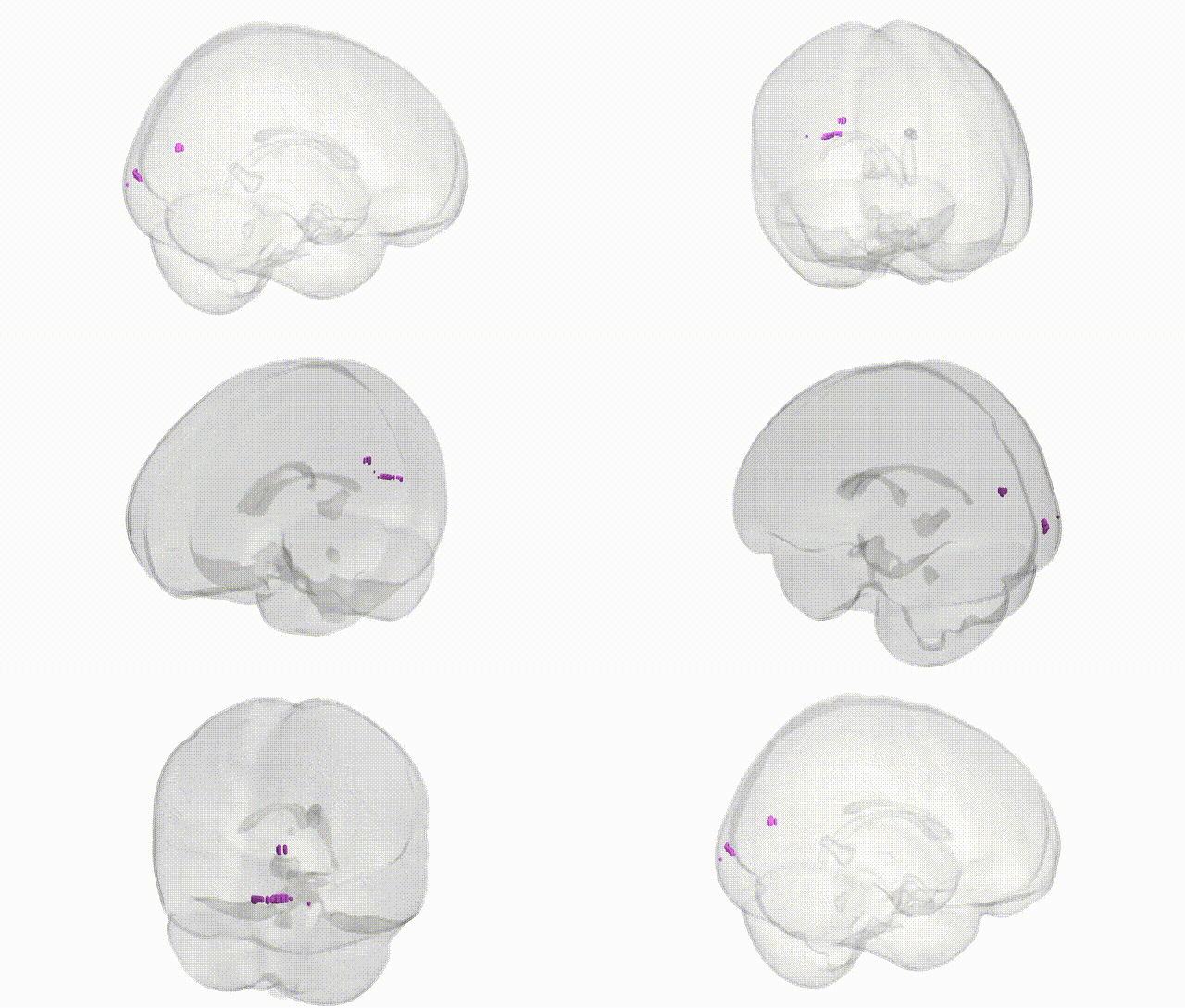
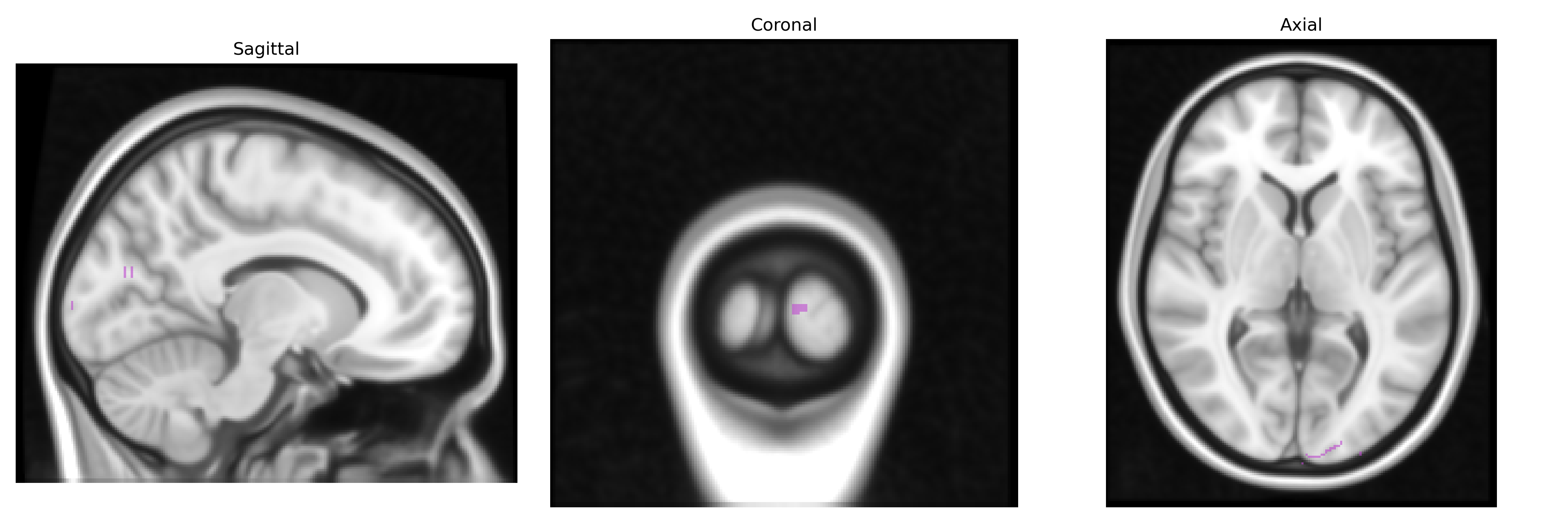
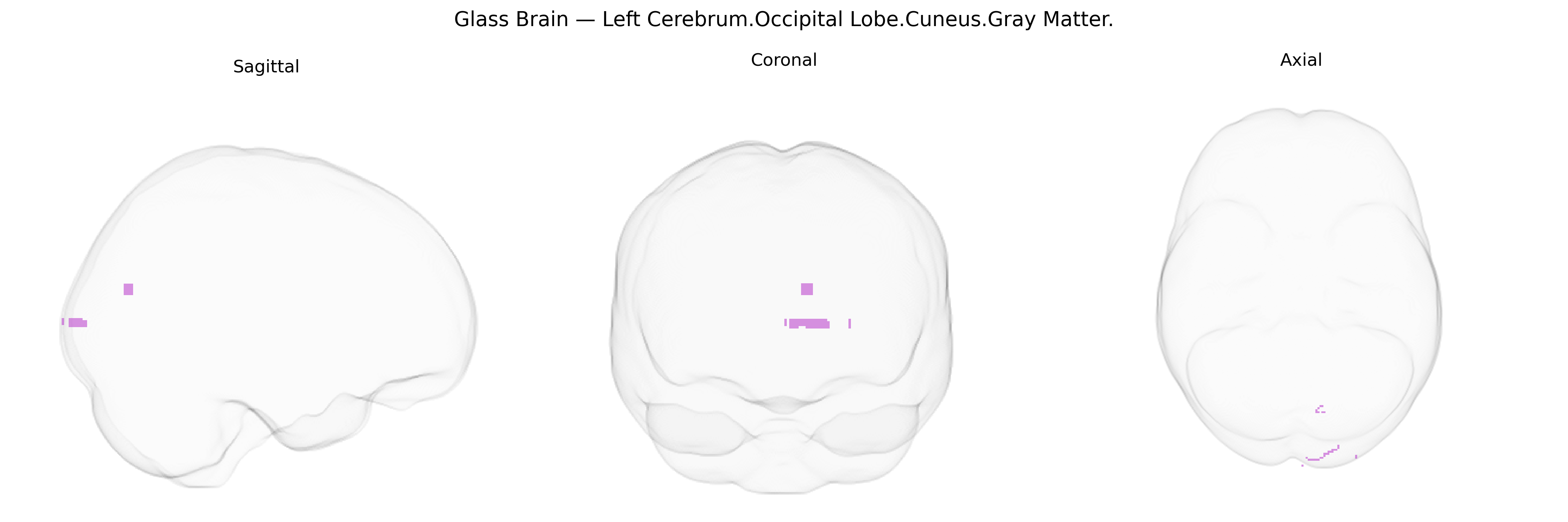
The bilateral Left Cerebrum.Occipital Lobe.Cuneus.Gray Matter corresponds to cortical tissue within the cuneus, a wedge-shaped region of the occipital lobe situated between the parieto-occipital sulcus and the calcarine fissure. This area forms part of the primary and associative visual cortex and is implicated in basic visual processing, including aspects of visual attention, visual imagery, and integration of visual information from the retinogeniculate pathways. Functionally, the cuneus contributes to the interpretation of motion, spatial relationships, and complex visual patterns, and shows connectivity with parietal and frontal regions involved in visuospatial cognition and eye-movement control.
Cuneus
The cuneus, a medial occipital gray-matter region involved in primary and higher-order visual processing as well as visuospatial and attentional functions, has emerged in imaging–genetics and GWAS studies as a heritable structure with polygenic influences spanning multiple neurobiological pathways; large-scale cortical thickness and surface-area GWAS (e.g., ENIGMA, UK Biobank) implicate common variants in genes regulating neurodevelopment, synaptic signaling, and myelination (such as MIR- and cadherin-related loci, although effects are generally regionally distributed rather than cuneus-specific), while twin and family studies show high heritability of cuneus morphology. Genetic associations involving the cuneus frequently appear in intermediate-phenotype analyses of psychiatric and neurological disorders: variants conferring risk for schizophrenia, major depressive disorder, bipolar disorder, autism spectrum disorder, and attention-deficit/hyperactivity disorder have been linked to altered cuneus gray-matter volume, thickness, or activity, particularly in visual and default-mode networks, and APOE-related risk for Alzheimer’s disease and other dementia-associated loci have been connected with cuneus atrophy or hypometabolism as part of posterior cortical involvement. GWAS of cognitive traits (e.g., general intelligence, educational attainment) and visual or visuospatial abilities often report polygenic effects on occipital and cuneus structure/function, while variants in genes related to visual pathway development (such as those affecting occipital lobe malformations or congenital blindness) can secondarily alter cuneus morphology; however, specific single-gene or locus effects uniquely targeting the bilateral cuneus are uncommon, and current evidence points to widespread, pleiotropic genetic architectures influencing this region in concert with broader visual and association cortices.
Overview generated by GPT-4o (2026).
Region ID: 533
Hemisphere: bilateral
Atlas: Talairach labels 1mm
Full Quality Version: Download MP4
Full Quality Version: Download MP4
Wali Sidiqyar*, Gaurav Rudravaram*, Elyssa M. McMaster, Trent M. Schwartz, Adam M. Saunders, Kurt G. Schilling, Bennett A. Landman "Introducing SPINS: A Shared Public Visualization Library of Neuroanatomical Structures." Medical Imaging with Deep Learning- short paper
This resource is licensed under CC0 1.0 Universal (Public Domain).