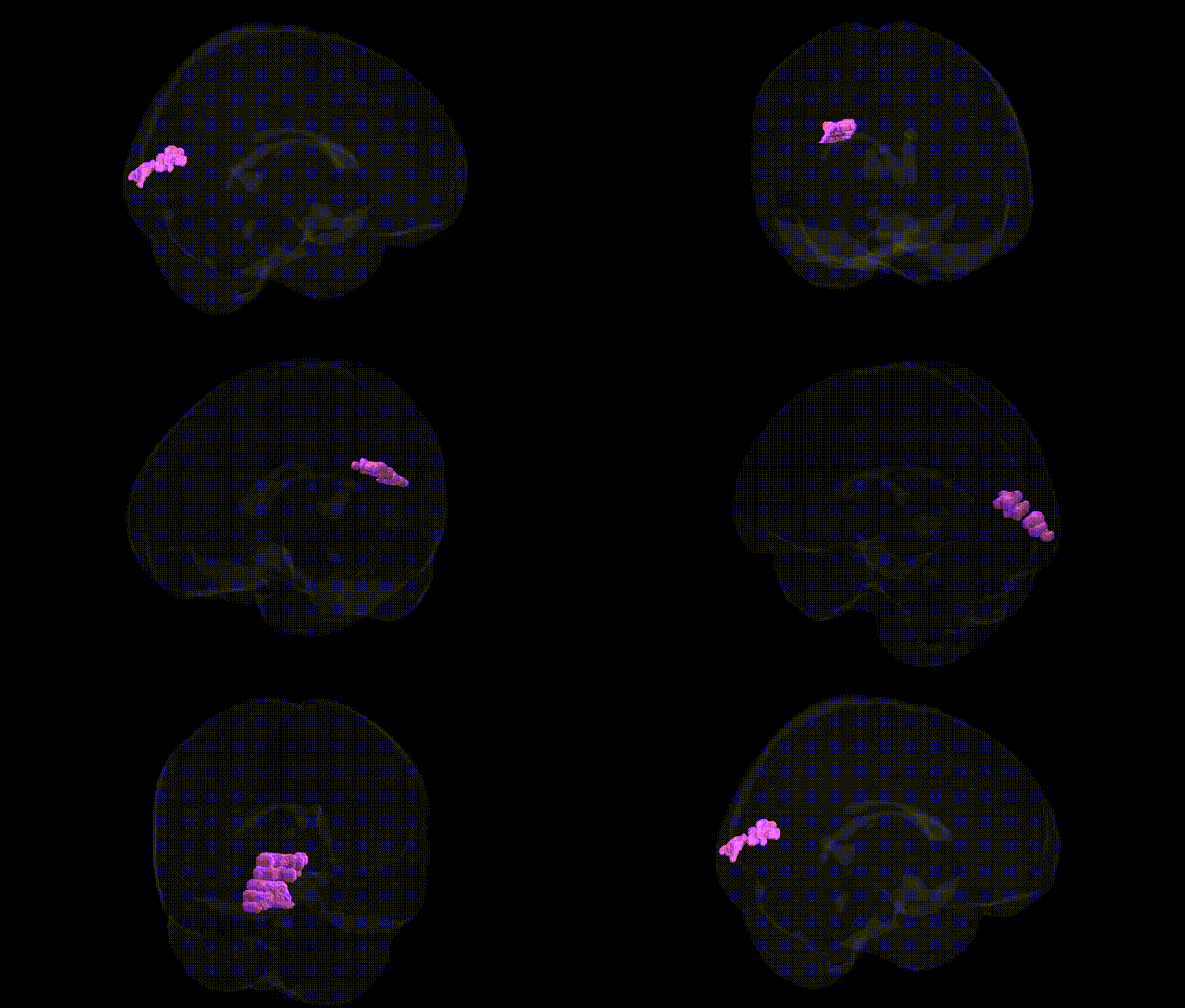
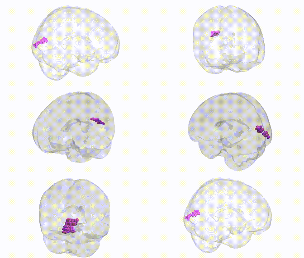
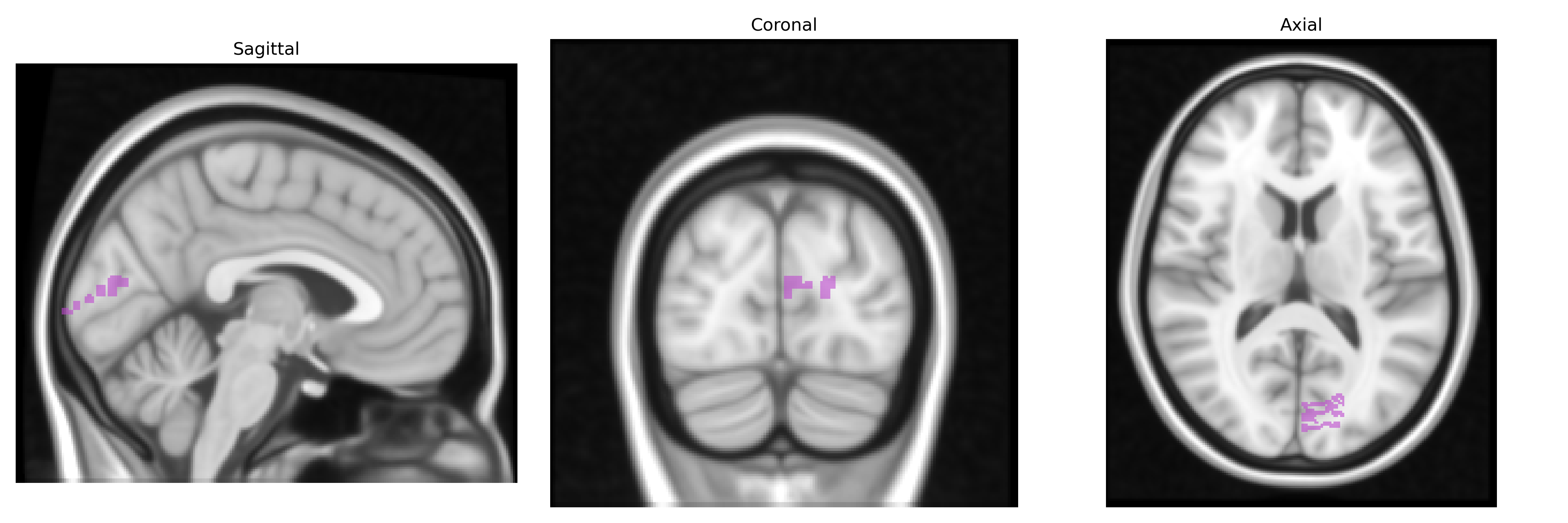
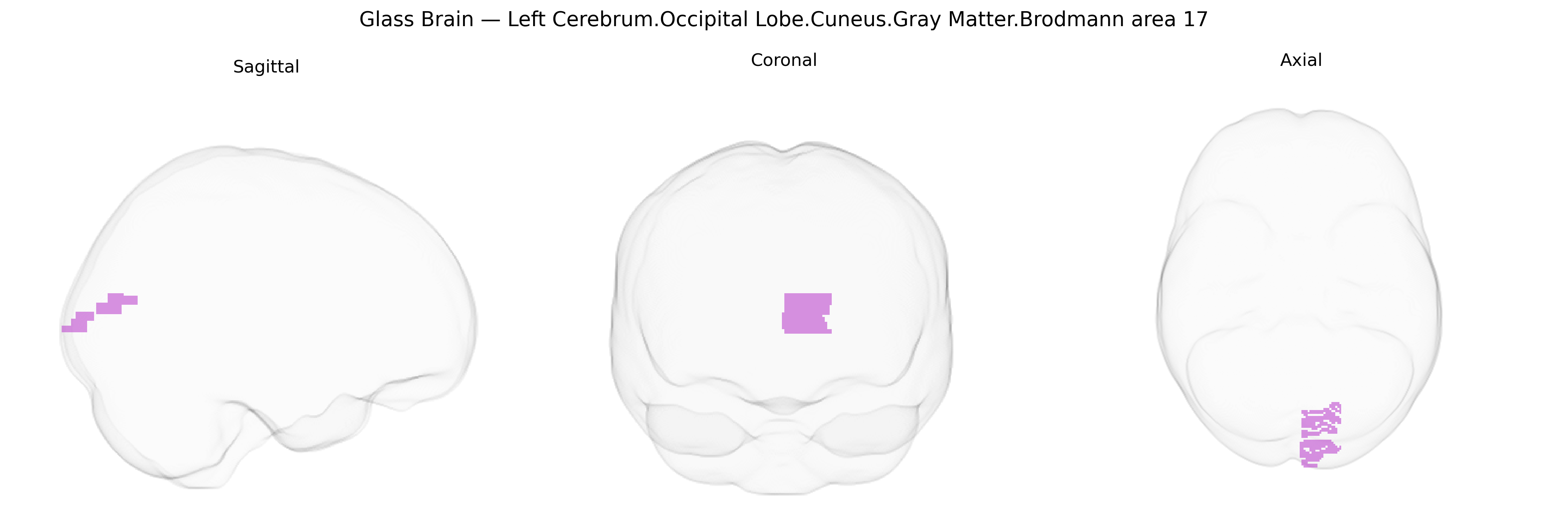
The bilateral Left Cerebrum.Occipital Lobe.Cuneus.Gray Matter.Brodmann area 17 corresponds to the primary visual cortex (V1) located in the medial aspect of the occipital lobe along the calcarine fissure, specifically within the cuneus region of the left hemisphere but considered here in a functionally bilateral context for visual processing. This area is the main cortical target of the lateral geniculate nucleus of the thalamus and contains a precise retinotopic map of the visual field, with a high density of small neurons and a distinctive laminar architecture, including the prominent line of Gennari in layer IV. Brodmann area 17 is critical for the initial stages of conscious visual perception, including edge detection, orientation, spatial frequency, and contrast, and serves as the primary input stage for higher-order visual association areas (BAs 18 and 19) involved in more complex processing of form, motion, and color. Brodmann area 17
The bilateral left cerebrum occipital lobe cuneus gray matter Brodmann area 17 (primary visual cortex) has been implicated in several genetic and GWAS-based associations, particularly through imaging-genetics studies that examine cortical thickness, surface area, and activation. Variants in genes involved in neurodevelopment and synaptic function (such as those in the Wnt, axon guidance, and neurotrophin pathways) have been associated with structural differences in BA17, often captured in large consortia like ENIGMA, which report heritable patterns of occipital cortical thickness and surface area linked to multiple loci across the genome. GWAS of visual cortex structure and function have identified associations with genes influencing ocular and visual pathway development, including loci near genes such as PAX6 and others related to eye morphology, although effects are typically polygenic and distributed rather than region-specific. Altered structure or activation of BA17 has also been reported in genetic studies of neuropsychiatric and neurodevelopmental conditions, including schizophrenia, bipolar disorder, autism spectrum disorder, and major depressive disorder, where risk variants in synaptic and glutamatergic genes correlate with occipital or primary visual cortex alterations. In migraine and visual aura, GWAS-implicated genes related to cortical excitability and ion channels overlap with regions encompassing BA17, consistent with imaging findings of altered visual cortex excitability. Additionally, rare variant and candidate gene studies in congenital blindness and albinism indicate that mutations affecting retinal and optic pathway development (e.g., in genes involved in melanin synthesis or retinal structure) indirectly drive reorganization and structural changes in BA17, linking specific genetic alterations to downstream changes in primary visual cortex organization.
Overview generated by GPT-4o (2026).
Region ID: 475
Hemisphere: bilateral
Atlas: Talairach labels 1mm
Full Quality Version: Download MP4
Full Quality Version: Download MP4
Wali Sidiqyar*, Gaurav Rudravaram*, Elyssa M. McMaster, Trent M. Schwartz, Adam M. Saunders, Kurt G. Schilling, Bennett A. Landman "Introducing SPINS: A Shared Public Visualization Library of Neuroanatomical Structures." Medical Imaging with Deep Learning- short paper
This resource is licensed under CC0 1.0 Universal (Public Domain).