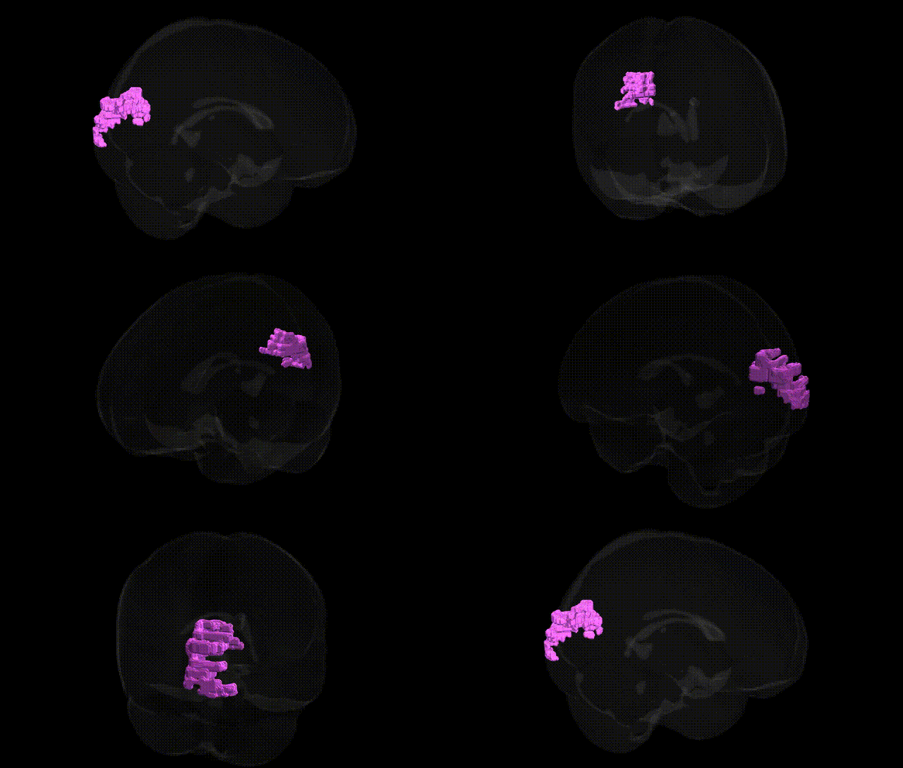
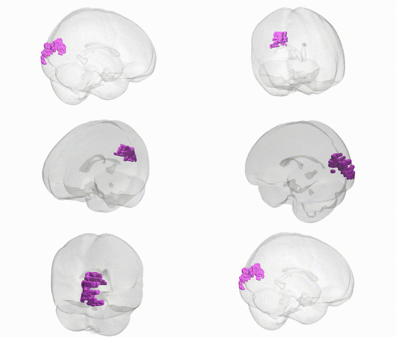
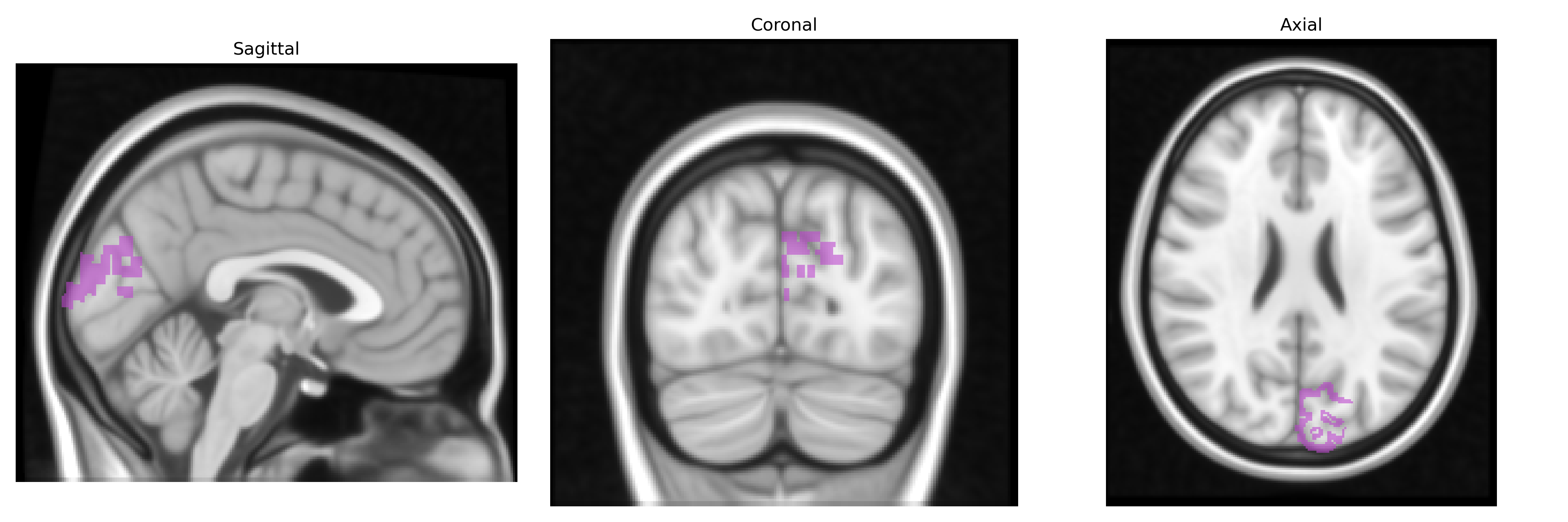
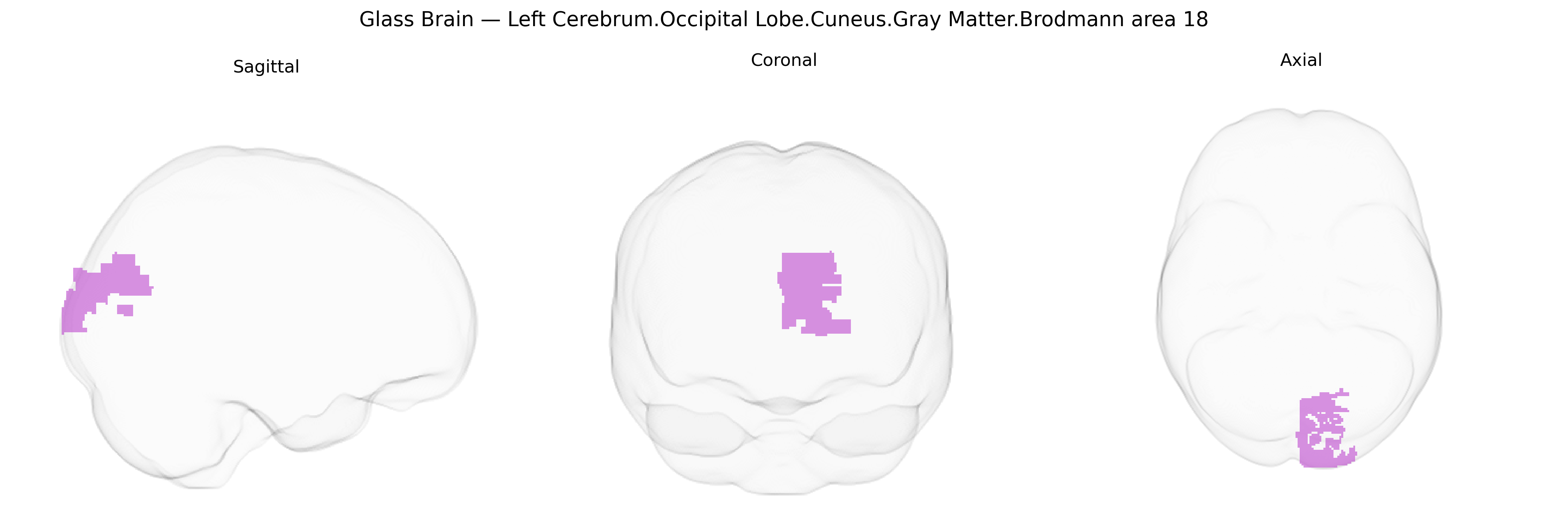
The bilateral Left Cerebrum.Occipital Lobe.Cuneus.Gray Matter.Brodmann area 18 corresponds to the secondary visual cortex (V2) located in the occipital lobe, immediately adjacent to the primary visual cortex (Brodmann area 17) along the banks of the calcarine fissure. This region receives strong feedforward input from V1 and projects to higher-order visual areas, contributing to early-stage processing of complex visual features such as orientation, spatial frequency, contour integration, stereoscopic depth, and simple forms of figure–ground segregation. Neurons in this area exhibit selectivity for binocular disparity and visual borders and participate in transforming retinotopic information into more abstract representations used in object recognition and visuospatial analysis. Functionally, damage or dysfunction in this region can contribute to deficits in visual discrimination and integration without complete loss of basic visual sensation. There is no direct link for this exact Talairach-defined label; a closely related structure is Brodmann area 18.
Genetic associations involving the bilateral left occipital cuneus gray matter in Brodmann area 18 (BA18) largely emerge from neuroimaging genetics and GWAS of brain structure, visual processing, and psychiatric or neurodevelopmental phenotypes. Variants in genes involved in synaptic development and plasticity (for example, BDNF, NRG1, and GRIN2B) and in axonal guidance or myelination (such as CNTNAP2 and MAG) have been linked to occipital cortical thickness or volume, including secondary visual cortex regions encompassing BA18, often in the context of imaging–genetics studies of schizophrenia, bipolar disorder, autism spectrum disorder, and major depression. Large-scale ENIGMA and UK Biobank GWAS of cortical thickness and surface area have identified multiple loci (e.g., near genes such as MCPH1, HMGA2, and microtubule- or chromatin-related genes) associated with occipital lobe morphometry, including cuneus/BA18 measures, with some loci overlapping those implicated in general cognitive ability, educational attainment, and intracranial volume. Additional GWAS of visual cortex structure and function have implicated genes involved in visual pathway development and retinal signaling in modulating occipital activation patterns, while GWAS of migraine (particularly with visual aura) and photosensitive epilepsy point to loci that affect excitability and network properties of posterior visual cortex, consistent with functional involvement of BA18. Overall, genetic influences on BA18 structure and function appear highly polygenic, overlapping with pathways for neurodevelopment, synaptic signaling, and cortical plasticity, and contribute to interindividual differences in visual processing, susceptibility to certain neuropsychiatric conditions, and structural brain endophenotypes.
Overview generated by GPT-4o (2026).
Region ID: 474
Hemisphere: bilateral
Atlas: Talairach labels 1mm
Full Quality Version: Download MP4
Full Quality Version: Download MP4
Wali Sidiqyar*, Gaurav Rudravaram*, Elyssa M. McMaster, Trent M. Schwartz, Adam M. Saunders, Kurt G. Schilling, Bennett A. Landman "Introducing SPINS: A Shared Public Visualization Library of Neuroanatomical Structures." Medical Imaging with Deep Learning- short paper
This resource is licensed under CC0 1.0 Universal (Public Domain).