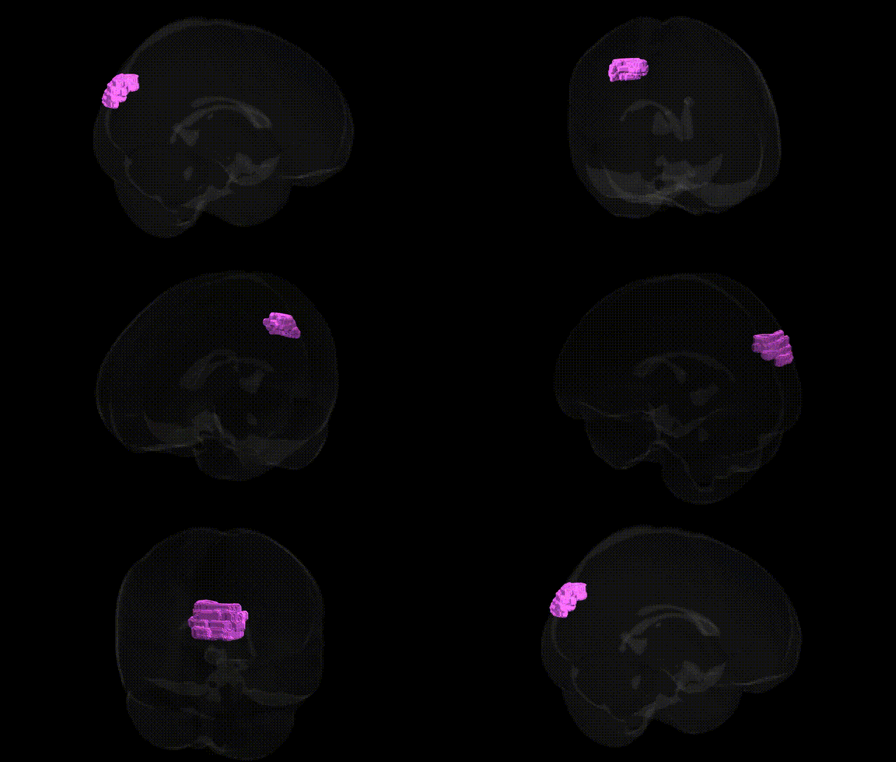
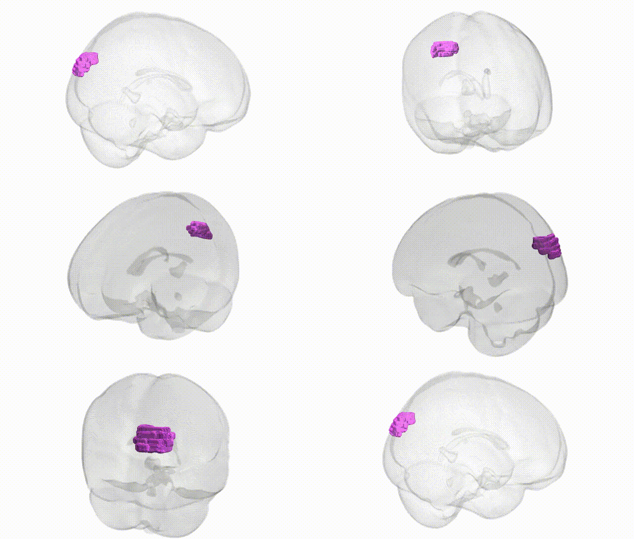
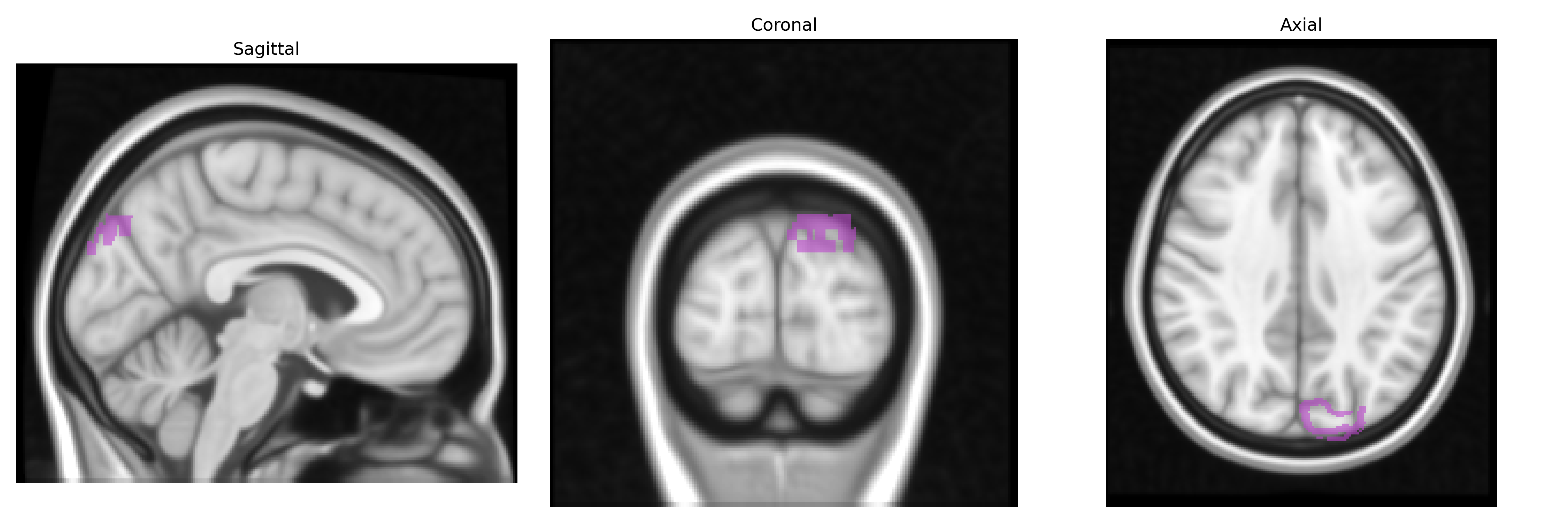
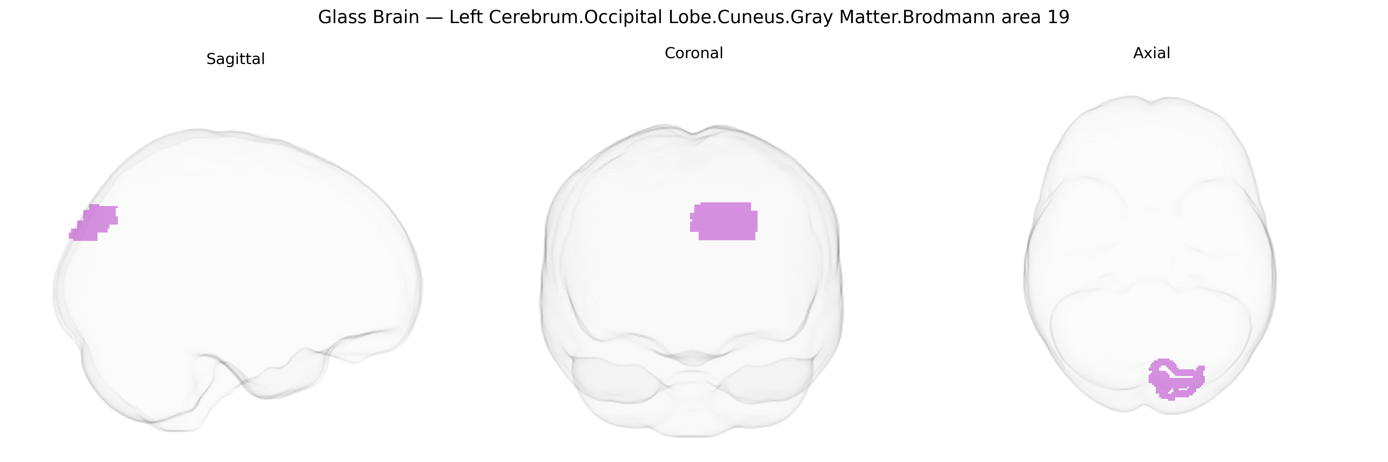
Bilateral Left Cerebrum.Occipital Lobe.Cuneus.Gray Matter.Brodmann area 19 corresponds to association visual cortex located in the dorsal medial occipital lobe, within the cuneus of the left hemisphere, extending across both sides relative to the midline representation in the Talairach 1 mm Atlas. Brodmann area 19 is part of extrastriate visual cortex and receives processed input from primary visual cortex (BA17) and secondary visual cortex (BA18), participating in higher-order visual functions such as complex form analysis, integration of motion and color information, and contributions to visual recognition and spatial processing. Neuronal populations here are organized in columnar and laminar patterns typical of neocortex, with dense corticocortical connections to other occipital, parietal, and temporal visual association areas, forming part of distributed networks for object perception and visuospatial cognition. There is no direct Wikipedia article for this exact Talairach label; a closely related structure is Brodmann area 19.
The bilateral left occipital BA19 (cuneus/visual association cortex) has been implicated indirectly in genetic studies of brain structure, cognition, and neuropsychiatric phenotypes, though few GWAS target this exact Talairach-defined label. Large imaging-genetics consortia (e.g., ENIGMA, UK Biobank) have identified common variants influencing occipital cortical thickness, surface area, and gray matter volume, frequently involving genes related to neurodevelopment, synaptic function, and neuronal migration, such as those in Wnt signaling (e.g., DACT1), axon guidance, and general cortical patterning pathways (e.g., variations near genes like PAX6 or LAMC3 in broader occipital cortex studies). Occipital and cuneus volume or activation patterns, including BA19, have been associated with genetic risk for schizophrenia, major depressive disorder, autism spectrum disorder, and ADHD via polygenic risk scores, as well as with visual perceptual skills, reading and dyslexia, and general cognitive performance, although the implicated variants typically act on distributed networks rather than BA19 specifically. Ophthalmologic and visual-function GWAS (e.g., for myopia, visual acuity, and optic nerve traits) sometimes show downstream effects on occipital visual areas, but the genetic signals are localized primarily to ocular and retinal genes (such as those near GJD2, RASGRF1) with secondary neural consequences. Overall, genetic associations involving this region are best characterized as part of broader occipital and visual network findings, with polygenic influences shaping its morphology and functional responsiveness rather than strong, region-specific single-gene effects.
Overview generated by GPT-4o (2026).
Region ID: 843
Hemisphere: bilateral
Atlas: Talairach labels 1mm
Full Quality Version: Download MP4
Full Quality Version: Download MP4
Wali Sidiqyar*, Gaurav Rudravaram*, Elyssa M. McMaster, Trent M. Schwartz, Adam M. Saunders, Kurt G. Schilling, Bennett A. Landman "Introducing SPINS: A Shared Public Visualization Library of Neuroanatomical Structures." Medical Imaging with Deep Learning- short paper
This resource is licensed under CC0 1.0 Universal (Public Domain).