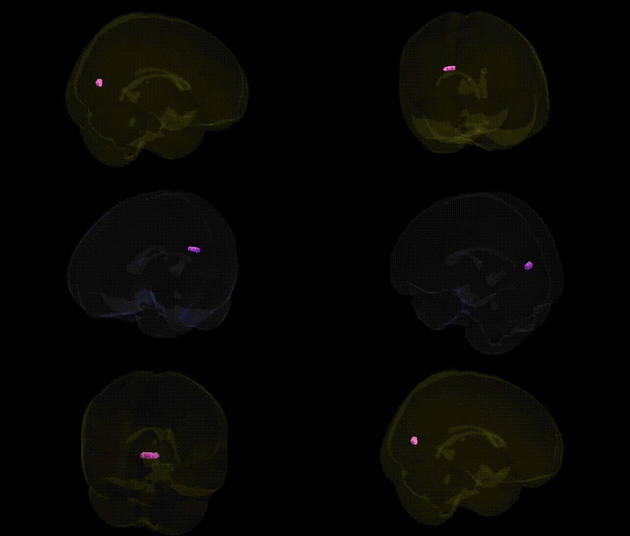
The bilateral Left Cerebrum.Occipital Lobe.Cuneus.Gray Matter.Brodmann area 23, as designated in the Talairach 1 mm atlas, refers to gray matter within the cuneus of the occipital lobe that falls within the stereotaxic territory commonly mapped to Brodmann area 23, a region traditionally classified as part of the ventral posterior cingulate cortex in the limbic lobe. Functionally, Brodmann area 23 is associated with visuospatial processing, episodic memory, self-referential thought, and integration of visual information with emotional and contextual cues, and it participates in large-scale networks such as the default mode network. In the Talairach labeling scheme, this assignment reflects an overlap between cytoarchitectonic fields and macroanatomical parcels, so the “cuneus” designation emphasizes its occipital location, whereas “Brodmann area 23” emphasizes its limbic/cingulate cytoarchitecture and connectivity profile. There is no direct Wikipedia article for this exact composite label; a closely related structure is the Posterior cingulate cortex.
The bilateral left cerebrum occipital lobe cuneus gray matter in the Talairach 1 mm Atlas, approximating Brodmann area 23 in posterior medial cortex/precuneus–cuneus territory, has been implicated in multiple genetic and GWAS findings through its role in visual processing, self-referential and default-mode network activity, and visuospatial integration. Large-scale imaging–genetics consortia (e.g., ENIGMA, UK Biobank) report SNP heritability for cortical thickness, surface area, and volume in the cuneus/precuneus, with common variants in genes related to neurodevelopment (e.g., BDNF, GRIN2B, DISC1, and synaptic/axonal guidance loci) showing associations with structural variation in this region. GWAS of brain imaging phenotypes identify significant loci influencing occipital and medial parietal morphometry (including cuneus) that overlap genes involved in neuronal differentiation, synaptic plasticity, and myelination, and these imaging endophenotypes often mediate genetic risk for psychiatric disorders such as schizophrenia, major depression, bipolar disorder, and autism spectrum disorder. Functional imaging–genetic studies link polymorphisms in serotonin transporter (SLC6A4), COMT, and APOE to altered activation or connectivity involving the cuneus/precuneus during memory, attention, and emotional tasks. Additionally, genetic risk variants for Alzheimer’s disease, Parkinson’s disease, migraine, and multiple sclerosis have been associated with altered structure or functional connectivity in this posterior medial cortex region, consistent with its vulnerability in neurodegeneration and its role in visual and default-mode networks, although no single gene has a specific, unique association limited to this exact Talairach-labeled subregion.
Overview generated by GPT-4o (2026).
Region ID: 706
Hemisphere: bilateral
Atlas: Talairach labels 1mm
Full Quality Version: Download MP4
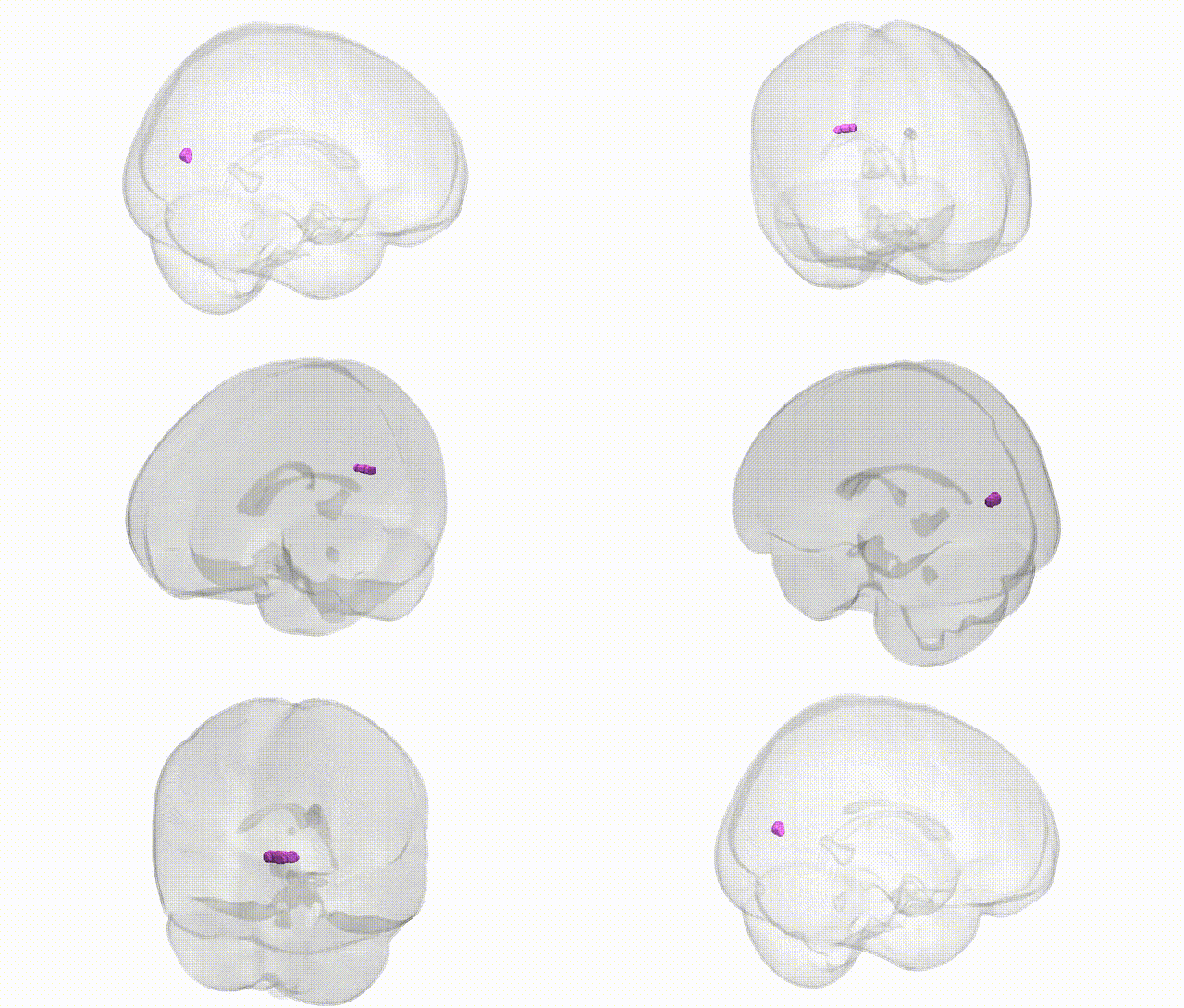
Full Quality Version: Download MP4
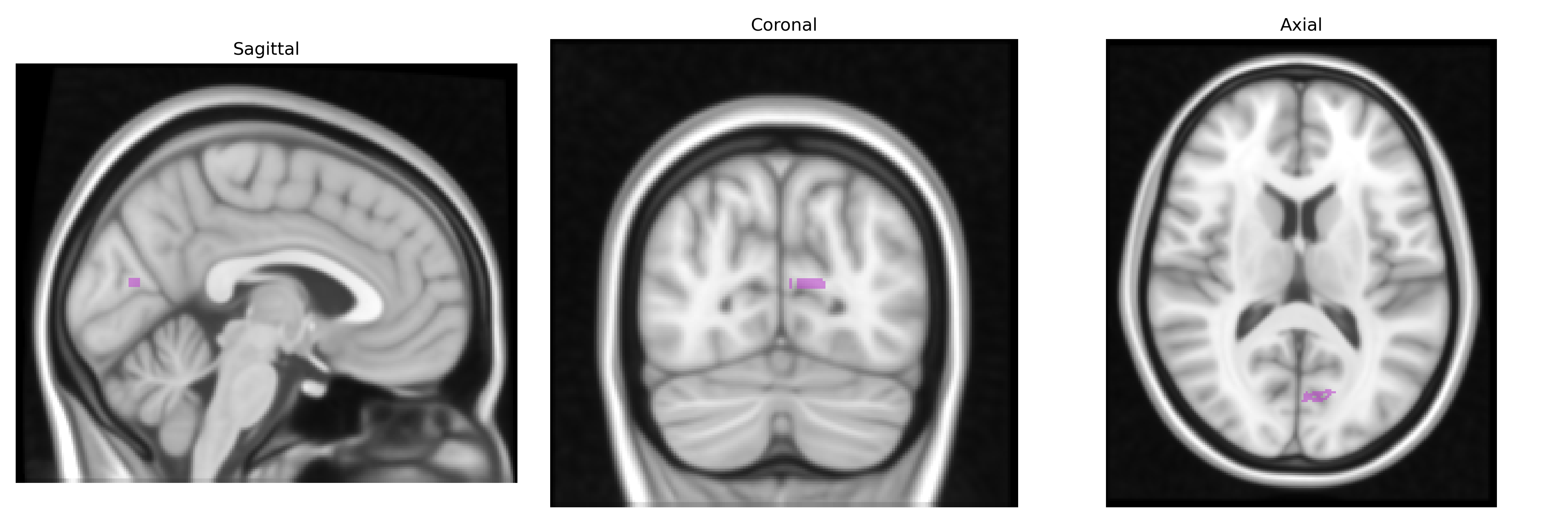
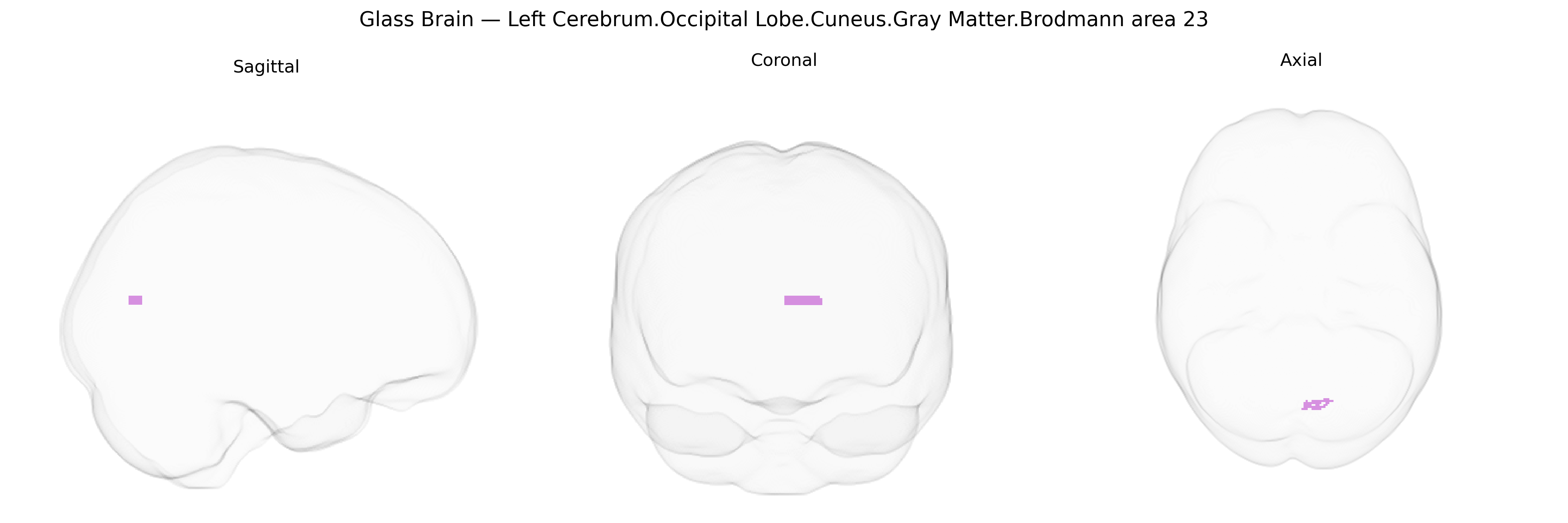
Wali Sidiqyar*, Gaurav Rudravaram*, Elyssa M. McMaster, Trent M. Schwartz, Adam M. Saunders, Kurt G. Schilling, Bennett A. Landman "Introducing SPINS: A Shared Public Visualization Library of Neuroanatomical Structures." Medical Imaging with Deep Learning- short paper
This resource is licensed under CC0 1.0 Universal (Public Domain).