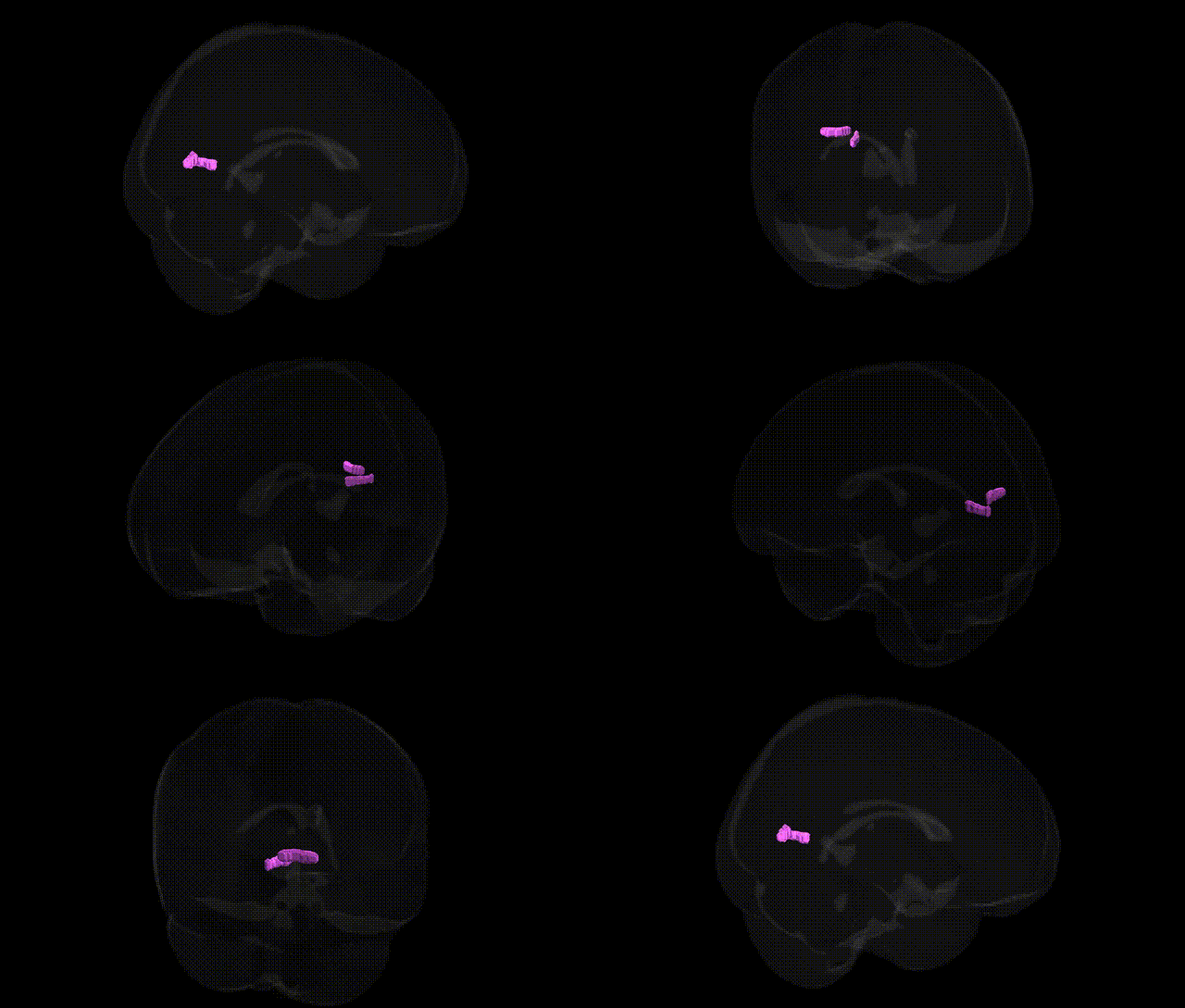
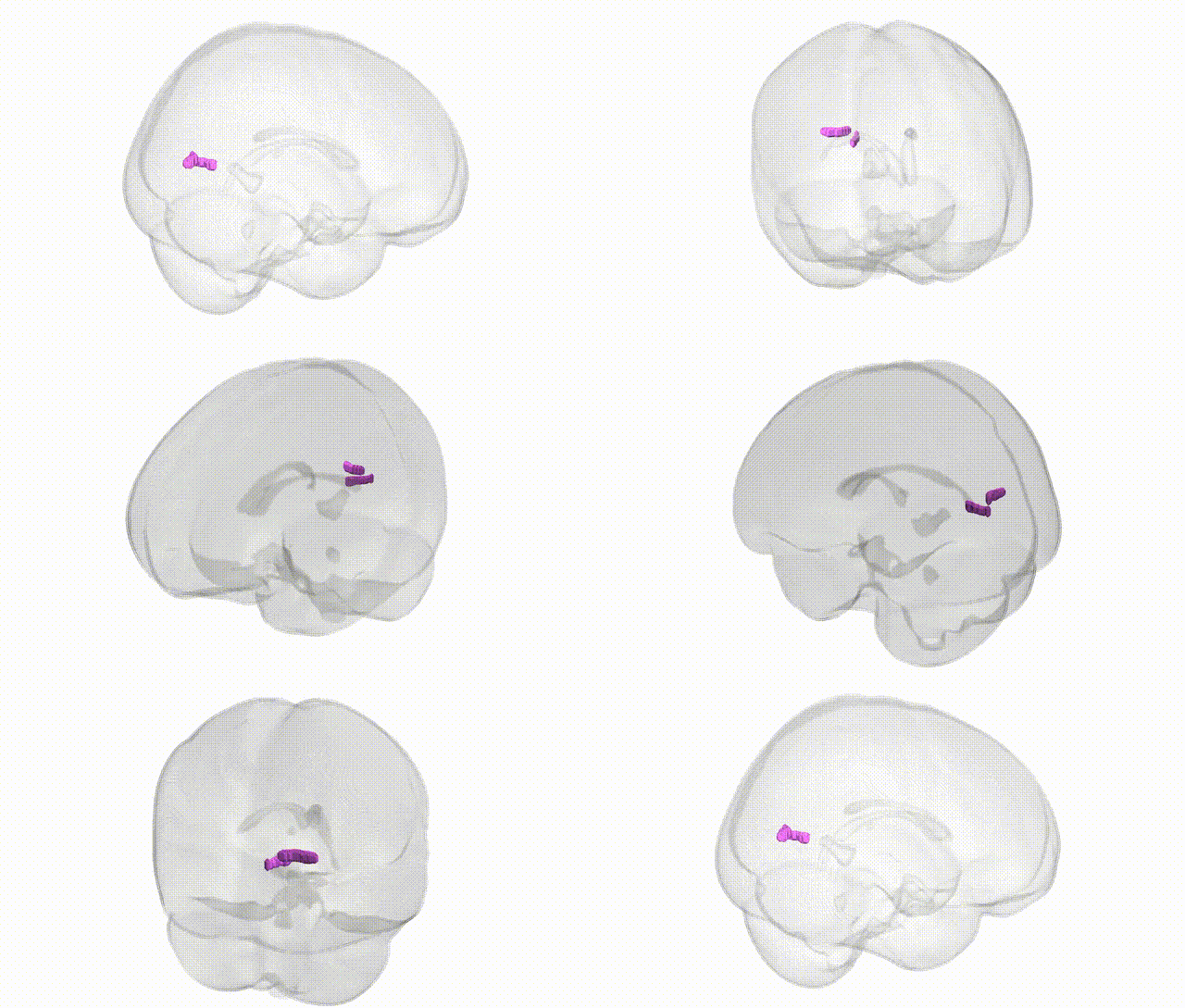
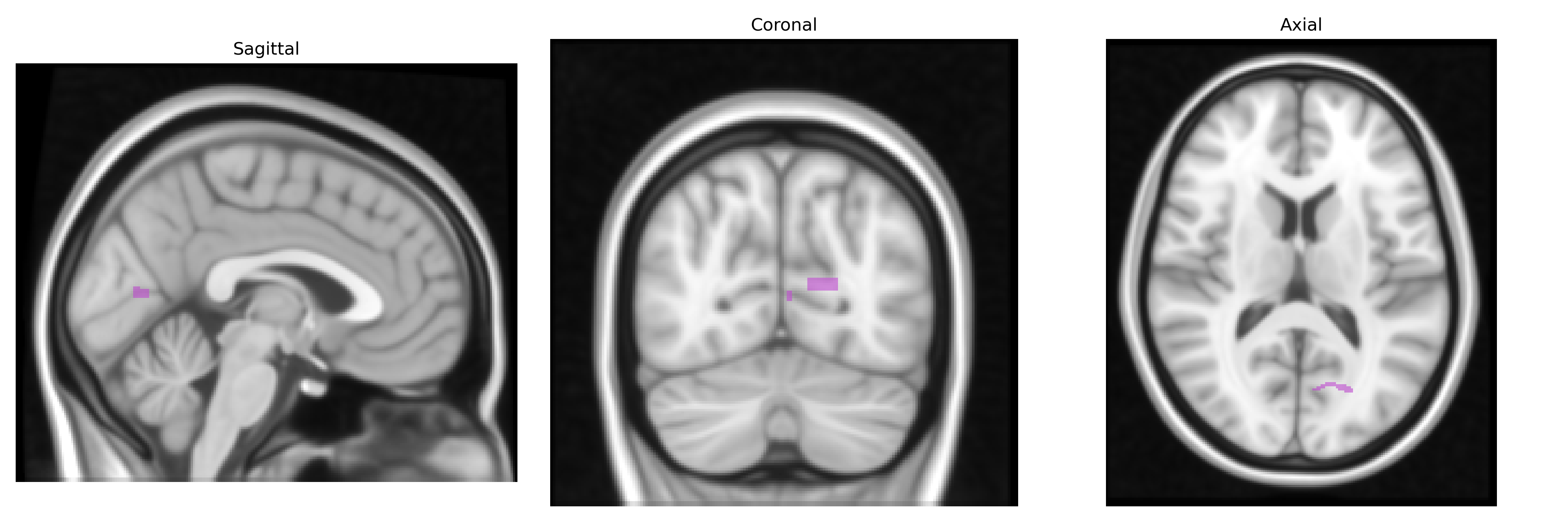
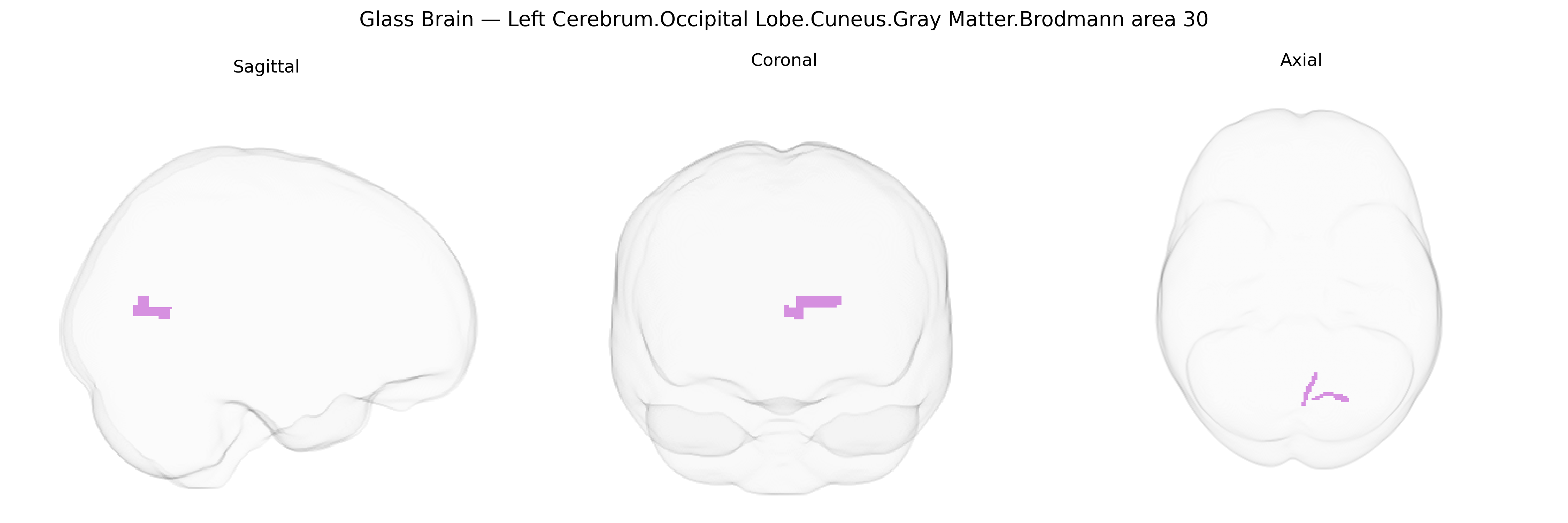
The bilateral Left Cerebrum.Occipital Lobe.Cuneus.Gray Matter.Brodmann area 30 corresponds to a portion of the retrosplenial cortex located in the medial occipital region, extending into the cuneus and bordering the parietal and limbic cortices. This area is associated with visuospatial processing, contextual and episodic memory, and integration of visual information with higher-order cognitive and limbic functions, often participating in networks involved in navigation and scene processing. Brodmann area 30 lies adjacent to other retrosplenial and posterior cingulate regions and shows strong connectivity with the hippocampal formation, parahippocampal cortex, and medial parietal areas, reflecting its role in memory-guided visual and spatial cognition. There is no direct link for Brodmann area 30 in the cuneus; a closely related structure is the Retrosplenial cortex.
The bilateral left cuneus in the occipital lobe (Brodmann area 30), part of medial visual cortex, has been implicated in several genetic and GWAS-based associations, though often indirectly through broader occipital or visual-network measures rather than BA30-specific loci. Large brain-imaging GWAS (e.g., ENIGMA and UK Biobank–based studies) have identified common variants in genes involved in neurodevelopment, synaptic function, and myelination—such as MAPT, KANSL1, WNT7B, and multiple loci near cell-adhesion and axon-guidance genes—that associate with occipital cortical thickness, surface area, or gray-matter volume encompassing the cuneus. Polygenic risk scores for schizophrenia, bipolar disorder, and major depressive disorder show correlations with structural or functional alterations in medial occipital regions including BA30, consistent with case–control imaging-genetics findings that link risk variants in genes like ZNF804A, CACNA1C, and NRGN to altered visual-network activation and connectivity. In neurodegenerative disease, APOE ε4 and other Alzheimer’s disease risk loci have been associated with atrophy patterns and hypometabolism extending to posterior cingulo-occipital and retrosplenial regions overlapping BA30. Additional associations involve visual processing traits and complex behaviors—such as reading ability, visuospatial performance, and resting-state visual-network connectivity—where common variants in neurodevelopmental and synaptic genes show small but detectable effects on cuneus morphology or activity, although most findings remain regionally coarse and not specific to Brodmann area 30 alone.
Overview generated by GPT-4o (2026).
Region ID: 660
Hemisphere: bilateral
Atlas: Talairach labels 1mm
Full Quality Version: Download MP4
Full Quality Version: Download MP4
Wali Sidiqyar*, Gaurav Rudravaram*, Elyssa M. McMaster, Trent M. Schwartz, Adam M. Saunders, Kurt G. Schilling, Bennett A. Landman "Introducing SPINS: A Shared Public Visualization Library of Neuroanatomical Structures." Medical Imaging with Deep Learning- short paper
This resource is licensed under CC0 1.0 Universal (Public Domain).