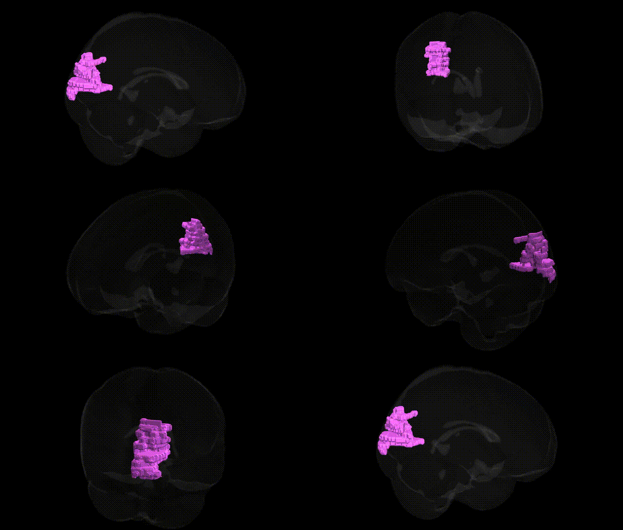
The bilateral Left Cerebrum.Occipital Lobe.Cuneus.White Matter corresponds to the subcortical myelinated fiber pathways underlying the cortical cuneus region of the occipital lobe, situated on the medial surface of the hemisphere between the calcarine sulcus and parieto‑occipital sulcus. These white matter tracts include feedforward and feedback connections linking primary and secondary visual cortices with higher-order visual and associative areas, contributing to the transmission and integration of visual information related to basic visual processing, spatial orientation, and aspects of visual attention. The cuneus white matter also participates in interhemispheric communication via callosal fibers and in cortico-subcortical loops with thalamic nuclei, supporting coordinated visual perception and visuospatial processing. No direct link exists specifically for the white matter portion; a related cortical region is described here: Cuneus.
The bilateral left cerebrum occipital lobe cuneus white matter, a visual association area involved in early-stage processing of visual information and integration with higher-order networks, has been implicated indirectly in genetic studies through imaging genetics and GWAS of brain structure, connectivity, and visual- and cognition-related traits rather than through region-specific loci. Large-scale GWAS of brain MRI measures (e.g., ENIGMA and UK Biobank) have identified polygenic influences on occipital cortical and adjacent white-matter volume, thickness, and microstructure, with variants in or near genes such as HMGA2, KIAA0586, and others contributing to global or regional brain size and white-matter integrity, which encompass occipital regions including the cuneus. Diffusion tensor imaging GWAS show that common variants affecting major posterior white-matter tracts (e.g., optic radiations and adjacent occipital fibers) are enriched in neurodevelopmental and axon guidance genes, some overlapping risk loci for schizophrenia, bipolar disorder, and major depression, suggesting that genetically driven alterations in occipital connectivity, including the cuneus white matter, may be part of broader neuropsychiatric risk architectures. Imaging genetics studies in conditions such as multiple sclerosis, Alzheimer’s disease, and migraine have demonstrated that disease-associated variants (e.g., in HLA, APOE, and TRPM8) can modulate lesion burden, atrophy, or functional activation patterns in occipital regions, but these effects are typically distributed and not specific to the cuneus white matter. Overall, current genetic evidence links this region to polygenic influences on visual system development, global white-matter organization, and vulnerability to complex neuropsychiatric and neurological disorders, rather than to discrete, cuneus-specific risk alleles.
Overview generated by GPT-4o (2026).
Region ID: 485
Hemisphere: bilateral
Atlas: Talairach labels 1mm
Full Quality Version: Download MP4
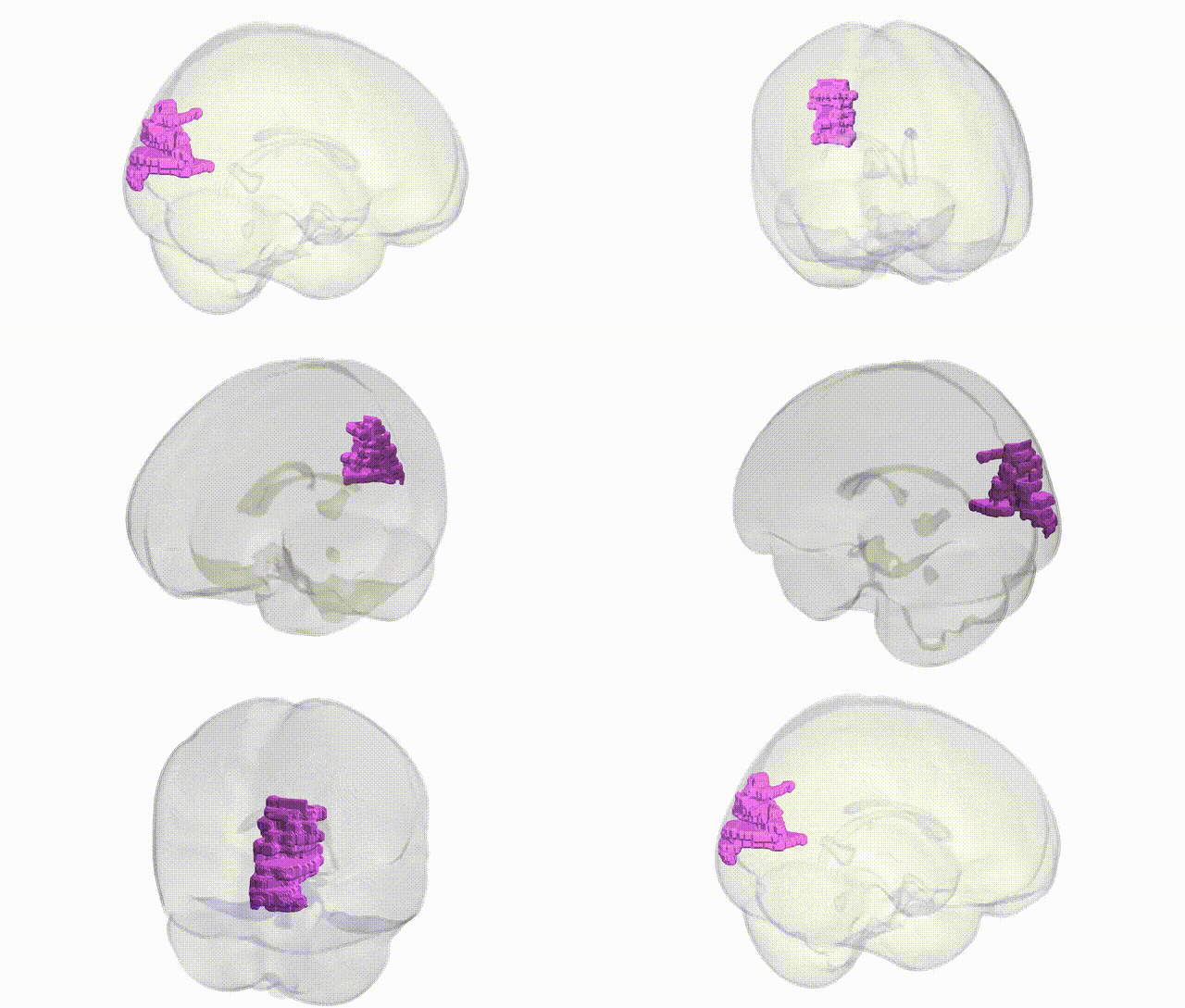
Full Quality Version: Download MP4
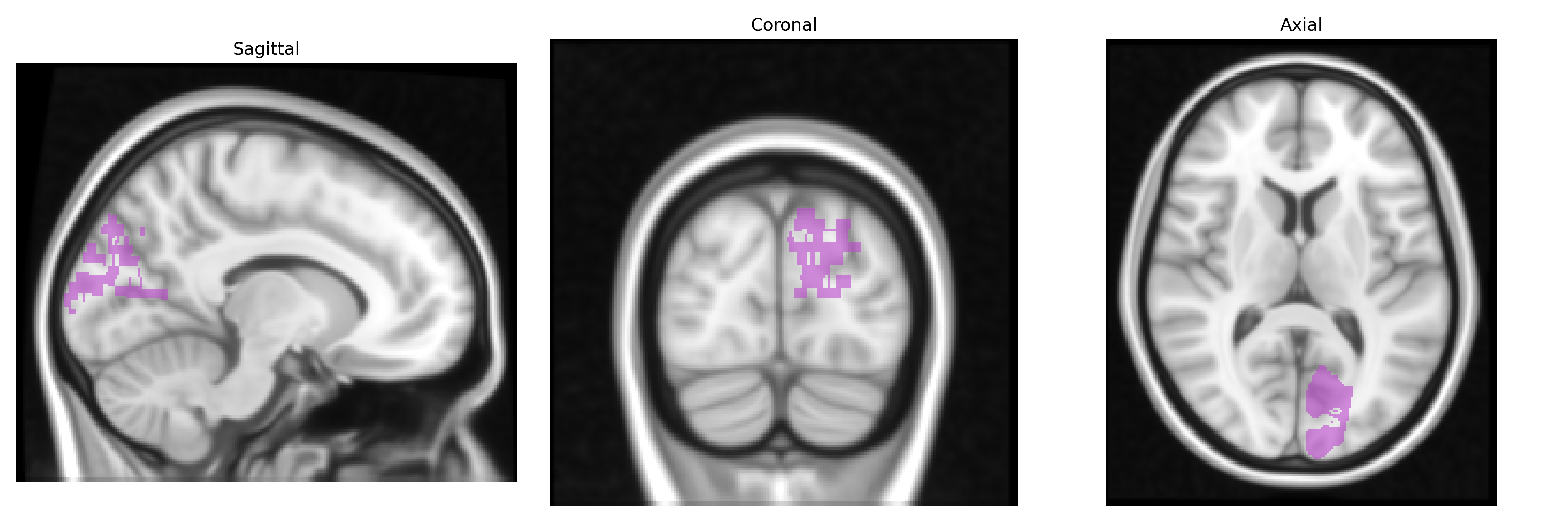
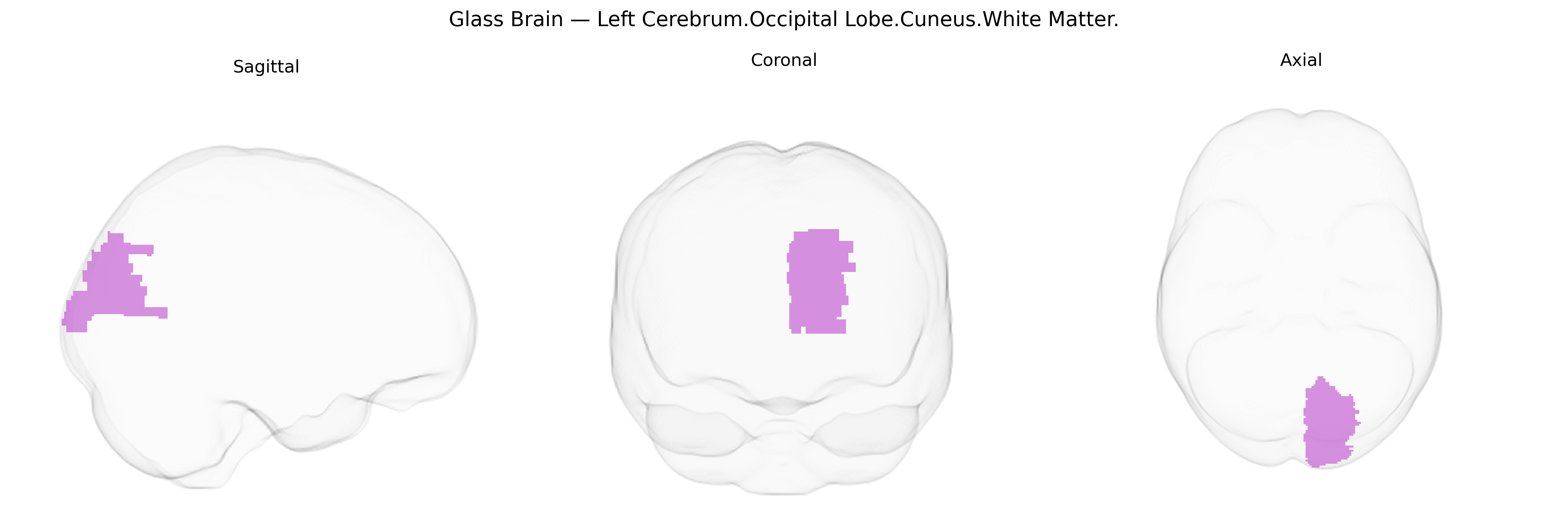
Wali Sidiqyar*, Gaurav Rudravaram*, Elyssa M. McMaster, Trent M. Schwartz, Adam M. Saunders, Kurt G. Schilling, Bennett A. Landman "Introducing SPINS: A Shared Public Visualization Library of Neuroanatomical Structures." Medical Imaging with Deep Learning- short paper
This resource is licensed under CC0 1.0 Universal (Public Domain).