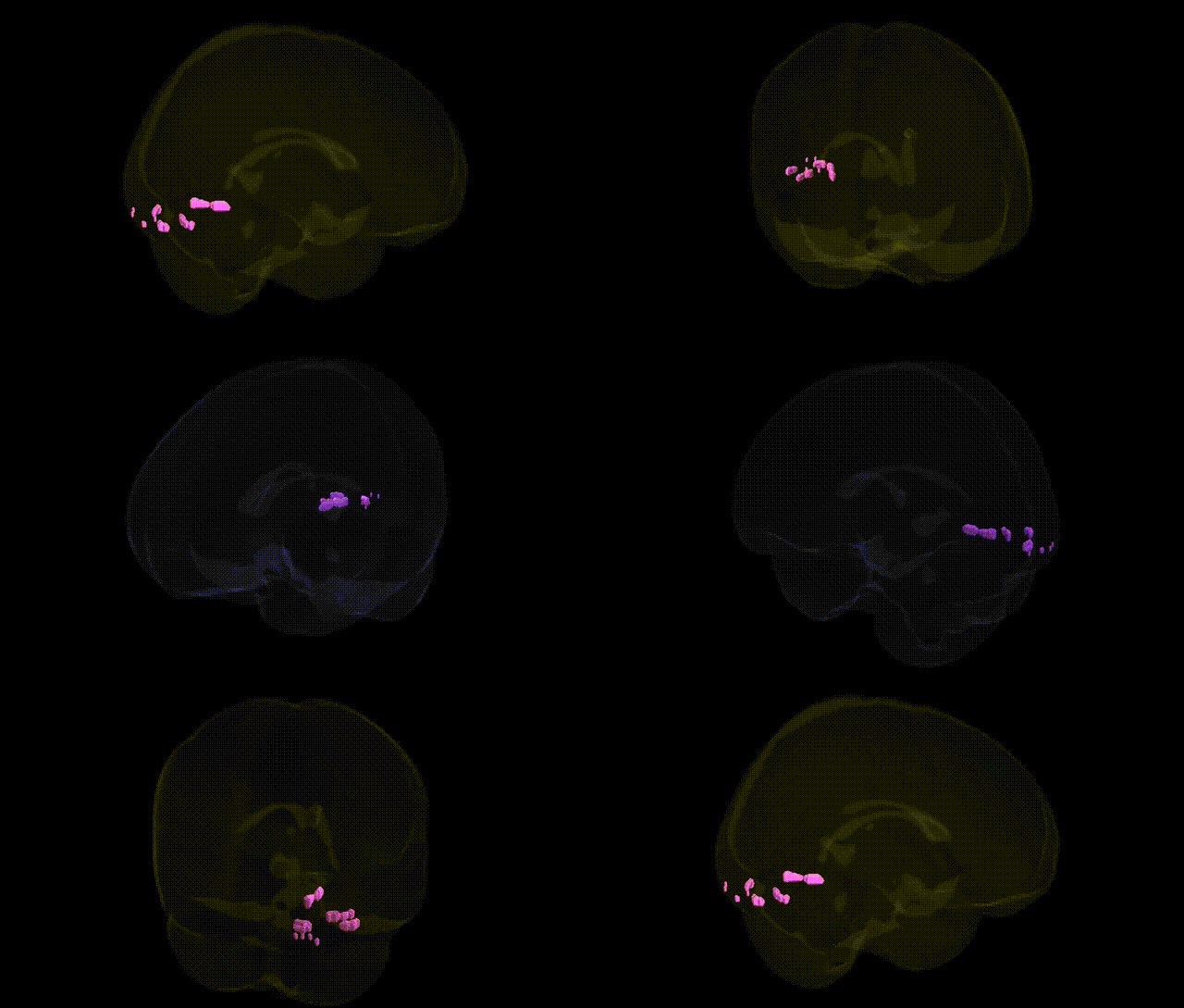
The bilateral Left Cerebrum.Occipital Lobe.Fusiform Gyrus corresponds to portions of the fusiform gyrus situated near or extending into the occipital lobe, forming part of the ventral visual processing stream. This cortical territory participates in high-level visual processing, including detailed analysis of complex shapes, faces, words, and category-specific objects, with functional specialization varying along its anterior–posterior axis. Inputs arise from earlier visual areas (such as V1–V4) and are integrated to support recognition and categorization, linking visual form information to memory and semantic systems. The fusiform gyrus exhibits hemispheric asymmetries, with left-sided regions particularly implicated in word and letter-form processing (e.g., the visual word form area), while overlapping or adjacent areas contribute to object and scene perception. There is no direct Wikipedia article for this exact Talairach label; see the related structure Fusiform gyrus.
The bilateral left cerebrum occipital lobe fusiform gyrus (often overlapping with occipitotemporal and “fusiform face area” territory) has been implicated in several genetic and GWAS-based associations, primarily through imaging-genetics and neuropsychiatric studies. Twin and heritability analyses show moderate to high genetic influence on fusiform gyrus volume, cortical thickness, and surface area, with common variants in pathways related to neurodevelopment (e.g., axon guidance, synaptic organization, and cell-adhesion genes) contributing to interindividual variability. Large imaging GWAS meta-analyses (such as ENIGMA and UK Biobank–based studies) report associations between fusiform/occipitotemporal morphology and variants in genes involved in neuronal migration and cortical patterning, though single loci often show small effects and limited reproducibility. Clinically, structural and functional alterations in fusiform and adjacent occipital regions have been linked to genetic risk for autism spectrum disorder, schizophrenia, bipolar disorder, and major depression, with polygenic risk scores for these conditions correlating with fusiform volume or activation differences. Rare monogenic or CNV syndromes affecting posterior cortical development (including 22q11.2 deletion and other neurodevelopmental CNVs) frequently show fusiform and occipitotemporal abnormalities, reflecting broader genetic disruption of cortical specialization. Additionally, genetic influences on face and word recognition abilities, dyslexia, and certain visual processing traits have been mapped to fusiform and occipitotemporal activation patterns, although specific risk loci for these cognitive phenotypes remain only partially characterized.
Overview generated by GPT-4o (2026).
Region ID: 202
Hemisphere: bilateral
Atlas: Talairach labels 1mm
Full Quality Version: Download MP4
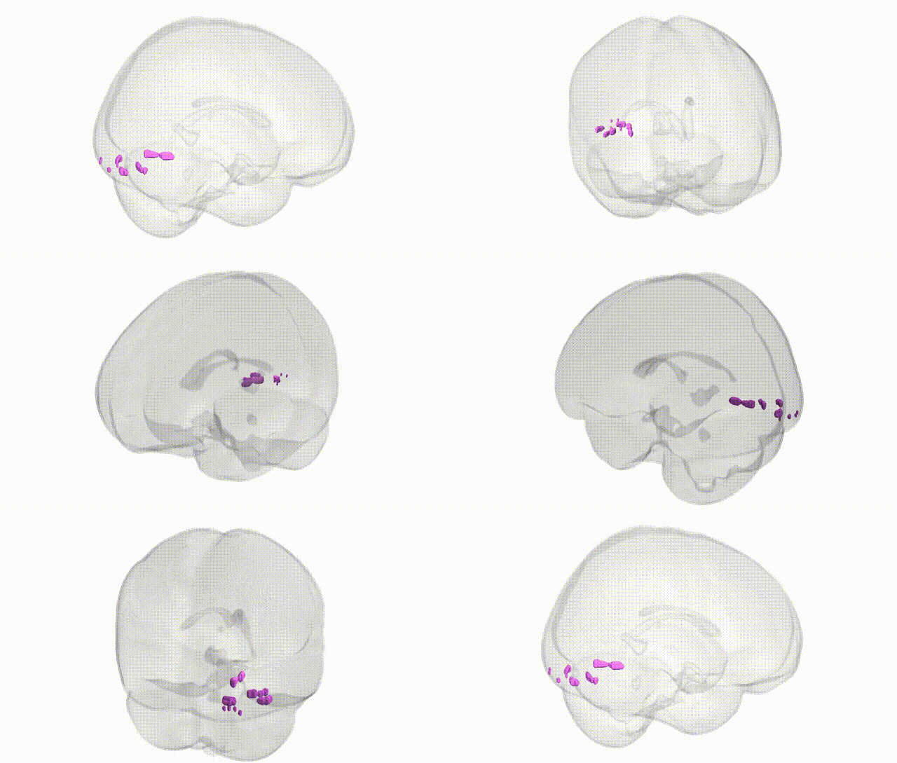
Full Quality Version: Download MP4
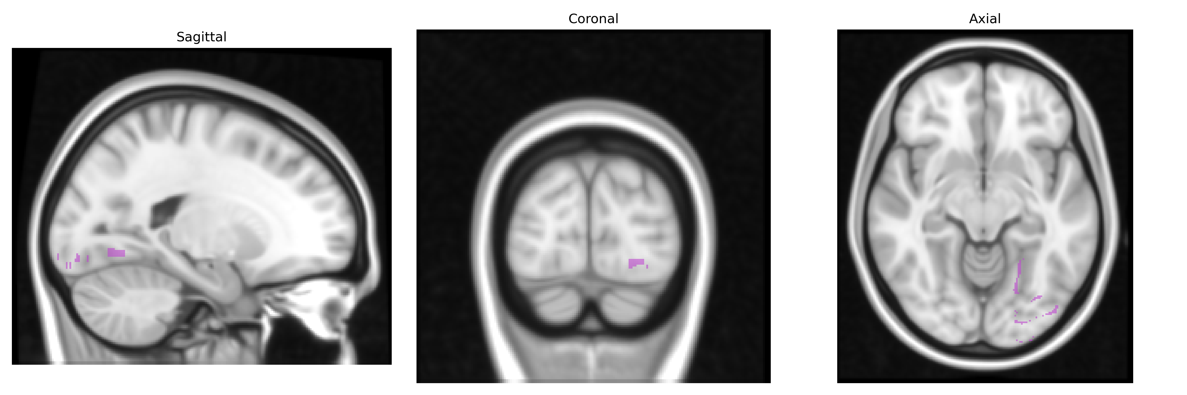
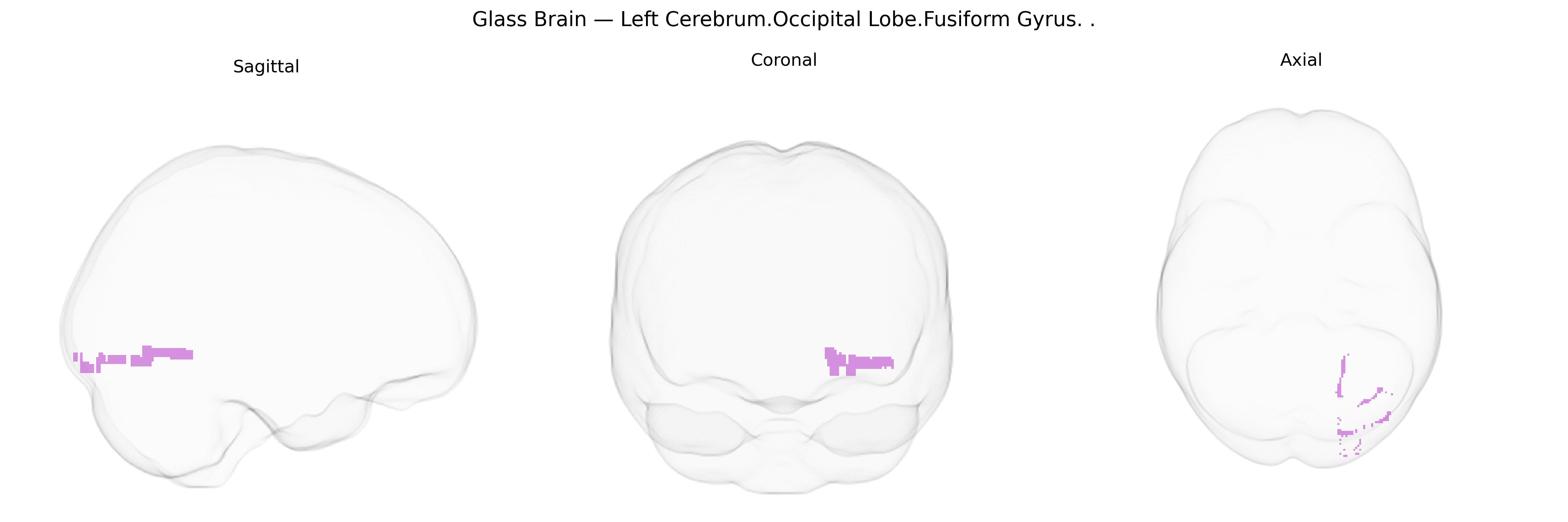
Wali Sidiqyar*, Gaurav Rudravaram*, Elyssa M. McMaster, Trent M. Schwartz, Adam M. Saunders, Kurt G. Schilling, Bennett A. Landman "Introducing SPINS: A Shared Public Visualization Library of Neuroanatomical Structures." Medical Imaging with Deep Learning- short paper
This resource is licensed under CC0 1.0 Universal (Public Domain).