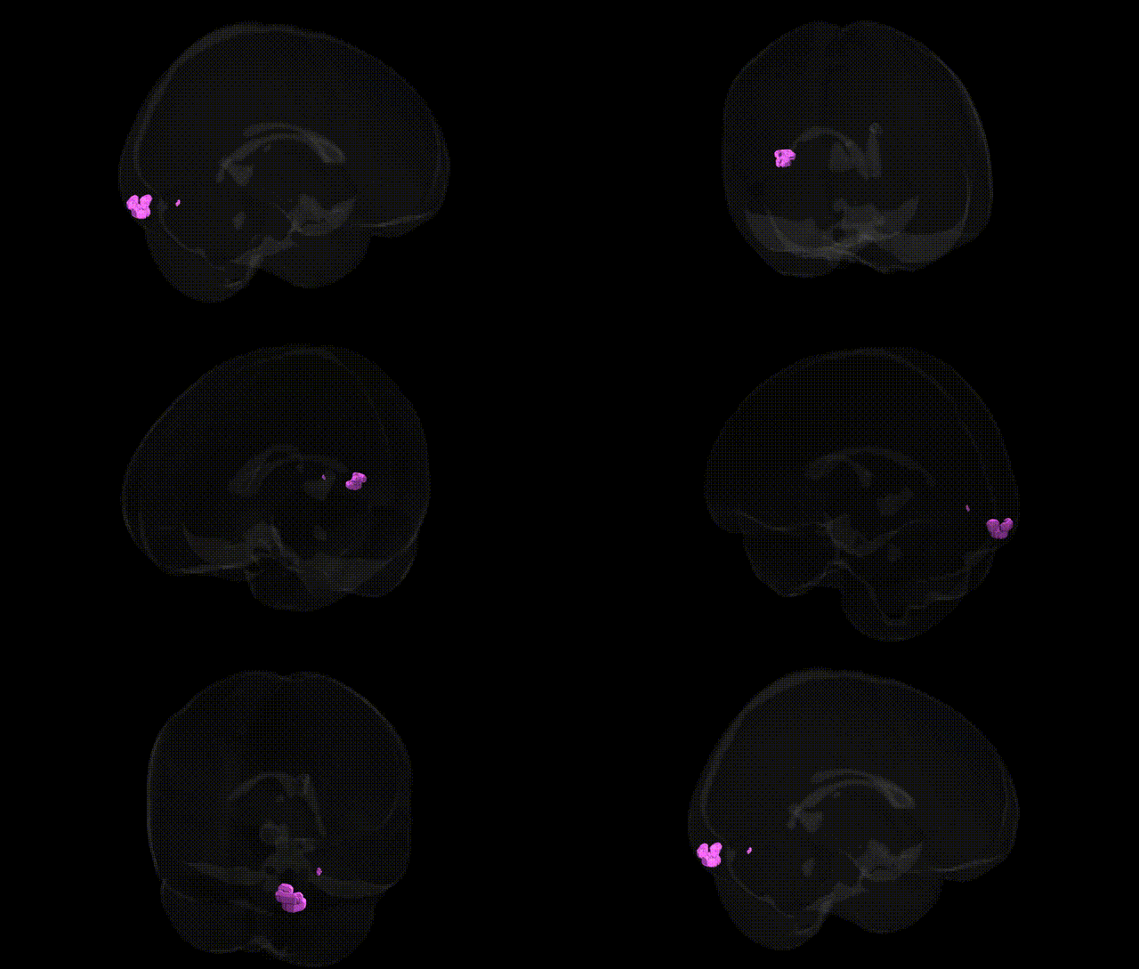
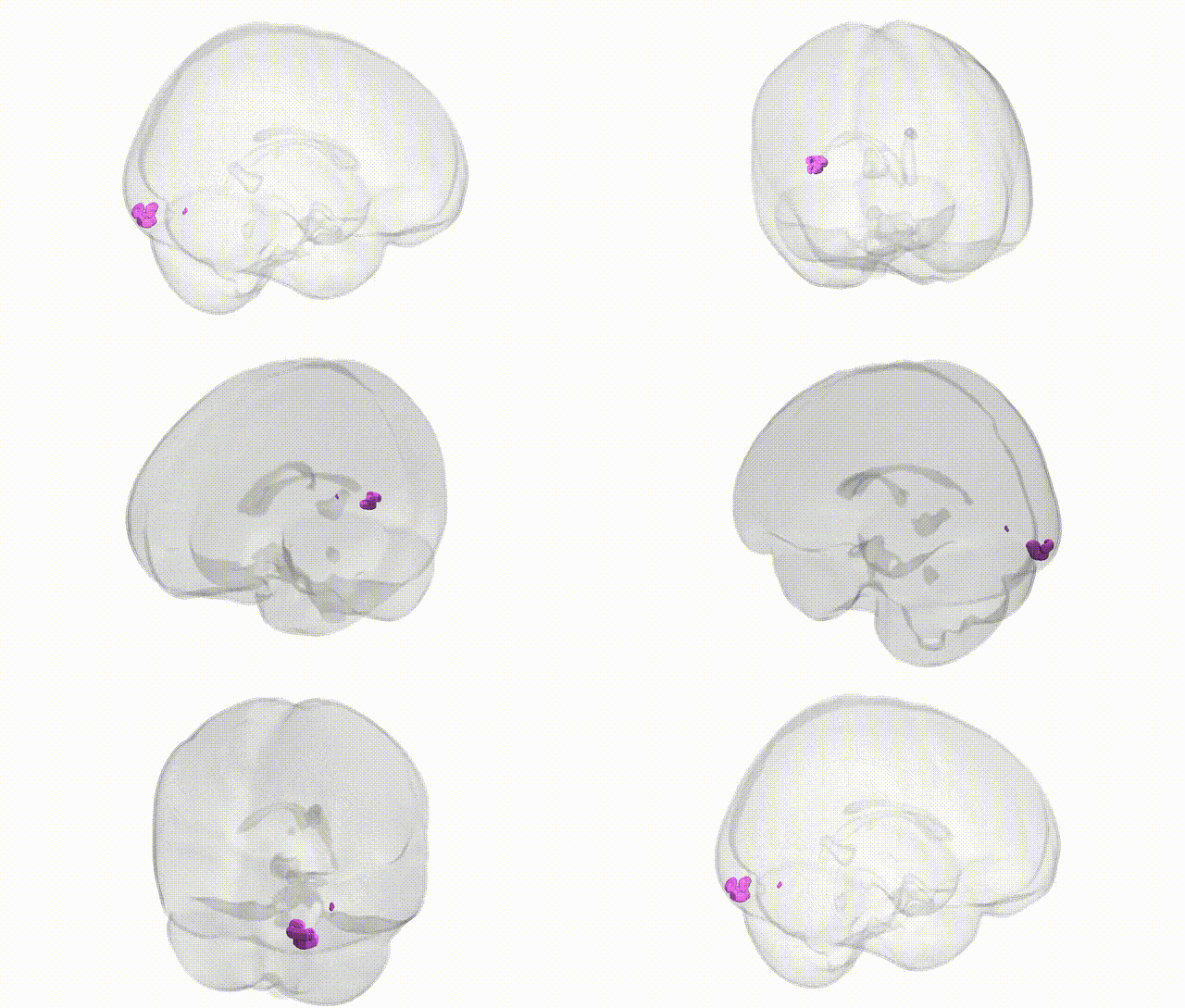
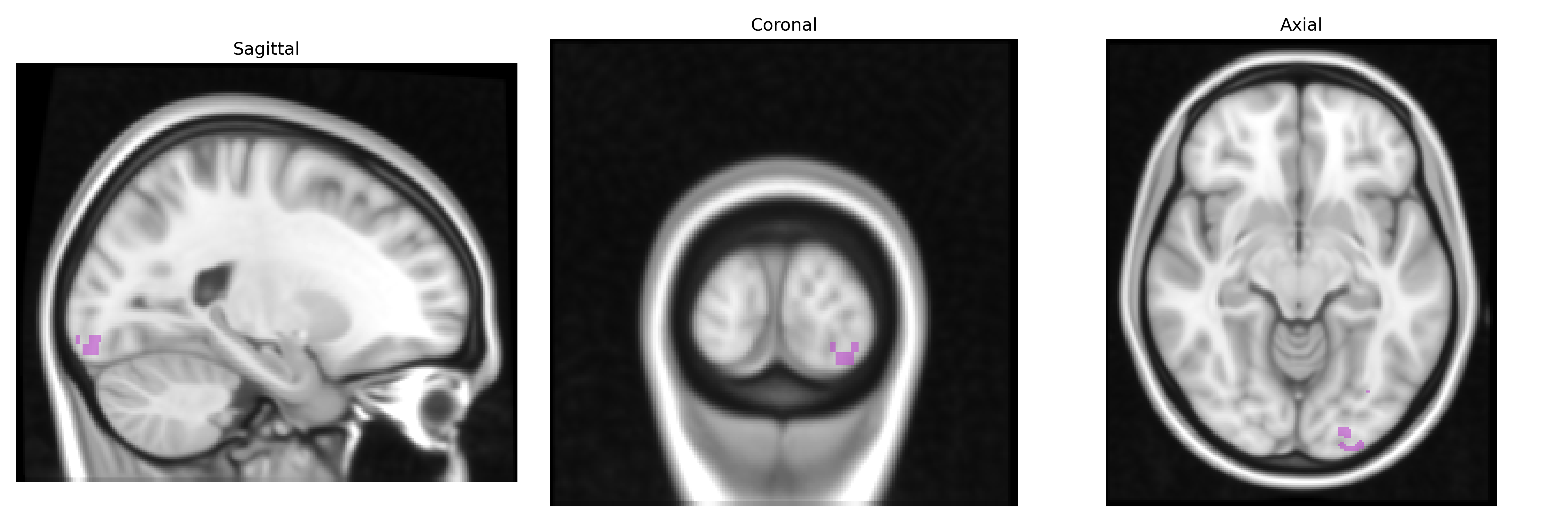
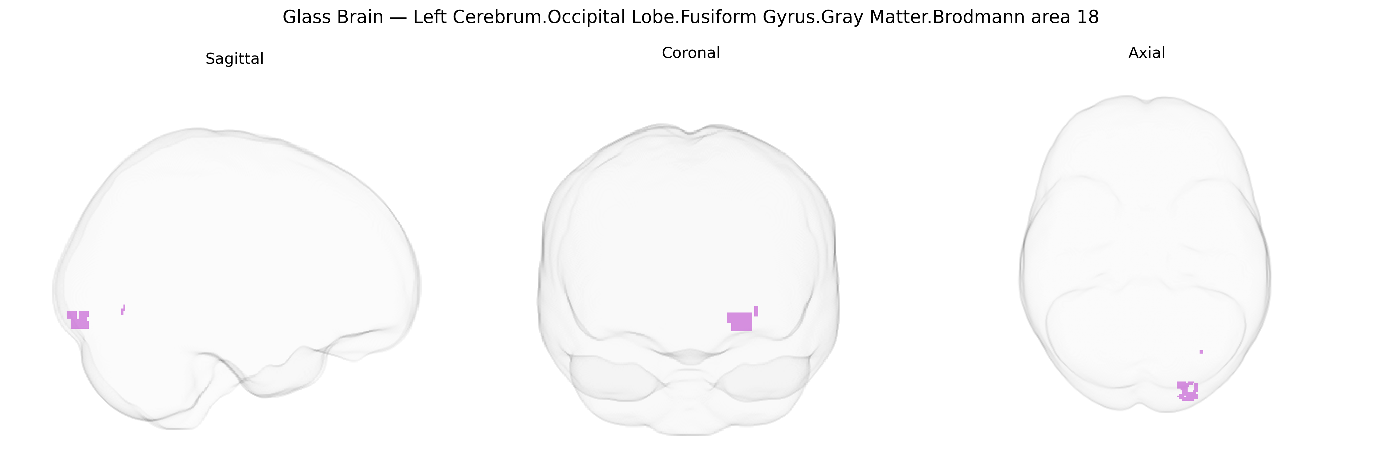
The bilateral Left Cerebrum.Occipital Lobe.Fusiform Gyrus.Gray Matter.Brodmann area 18 corresponds to secondary visual cortex (V2) localized within the fusiform gyrus of the occipital lobe, representing a portion of extrastriate visual cortex involved in early-stage processing of complex visual information such as contours, orientation, and spatial relationships. As part of Brodmann area 18, this region receives strong feedforward input from primary visual cortex (V1, BA17) and distributes processed visual signals to higher-order visual association areas, participating in the integration of form, texture, and pattern features that support object recognition and visual perception. Histologically, BA18 is characterized by a well-developed granular layer IV, dense myelination, and cytoarchitectonic features typical of visual association cortex, and functionally it contributes to contextual modulation of visual signals and the refinement of visual representations along the ventral visual stream. There is no direct Wikipedia article for this exact Talairach-defined region; a closely related structure is Brodmann area 18.
The bilateral left cerebrum occipital lobe fusiform gyrus gray matter in Brodmann area 18 (a visual association cortex region adjacent to BA19 and ventral temporal fusiform areas) has been implicated in several genetic and GWAS-based findings, largely through imaging genetics rather than region-specific single-gene studies. Polygenic influences on occipital cortical thickness and surface area, including BA18/secondary visual cortex, have been identified in large neuroimaging GWAS consortia such as ENIGMA, with common variants in loci near genes involved in neurodevelopment and synaptic function (e.g., variants near PAX6, TBR1, and cell-adhesion/synaptogenesis genes) contributing modestly to structural variation. Although fusiform gyrus genetic work often focuses more anteriorly (e.g., face-selective fusiform and word-form areas), BA18/occipital fusiform segments show heritable variation in volume and activity tied to visual processing performance, with twin and GWAS studies linking polygenic scores for general cognitive ability, educational attainment, and reading-related traits to structural and functional measures in this region. Imaging genetics in neuropsychiatric disorders suggests that schizophrenia, bipolar disorder, and major depression risk variants exert small effects on occipital/fusiform gray matter and visual processing networks, while autism spectrum disorder risk loci (including synaptic and chromatin-remodeling genes such as SHANK3, CHD8, and SCN2A) have been associated with atypical activation and connectivity across occipito-temporal visual regions, including BA18-adjacent fusiform cortex, during face and social perception tasks. Additionally, GWAS of migraine and visual aura, as well as polygenic risk for occipital lobe epilepsy, implicate genetic influences on excitability and cortical visual network structure that encompass BA18, though findings are typically network-level rather than specific to this Talairach-defined subregion. Overall, genetic associations for this bilateral BA18 fusiform/occipital region are polygenic, small in effect, and primarily observed through imaging-genetic correlations with visual, cognitive, and neuropsychiatric traits rather than through single, robustly replicated locus–region mappings.
Overview generated by GPT-4o (2026).
Region ID: 203
Hemisphere: bilateral
Atlas: Talairach labels 1mm
Full Quality Version: Download MP4
Full Quality Version: Download MP4
Wali Sidiqyar*, Gaurav Rudravaram*, Elyssa M. McMaster, Trent M. Schwartz, Adam M. Saunders, Kurt G. Schilling, Bennett A. Landman "Introducing SPINS: A Shared Public Visualization Library of Neuroanatomical Structures." Medical Imaging with Deep Learning- short paper
This resource is licensed under CC0 1.0 Universal (Public Domain).