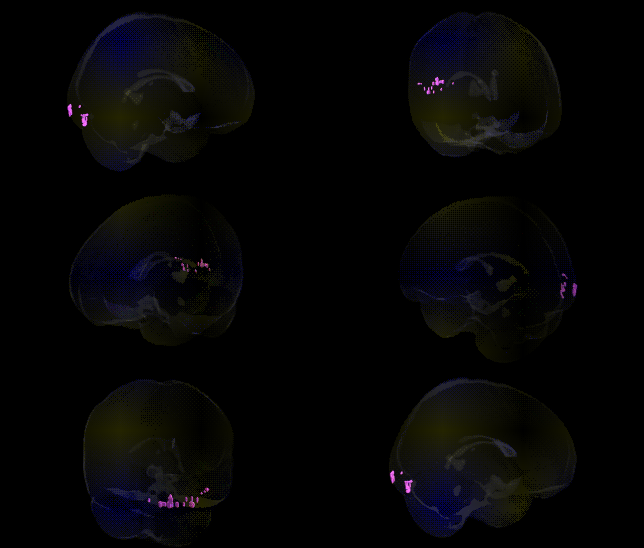
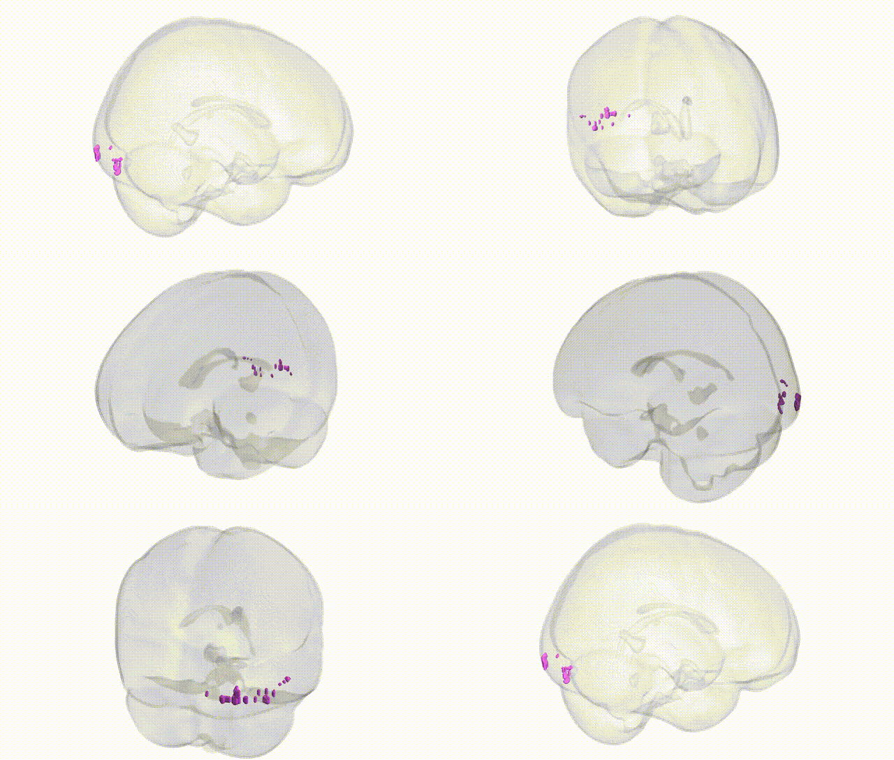
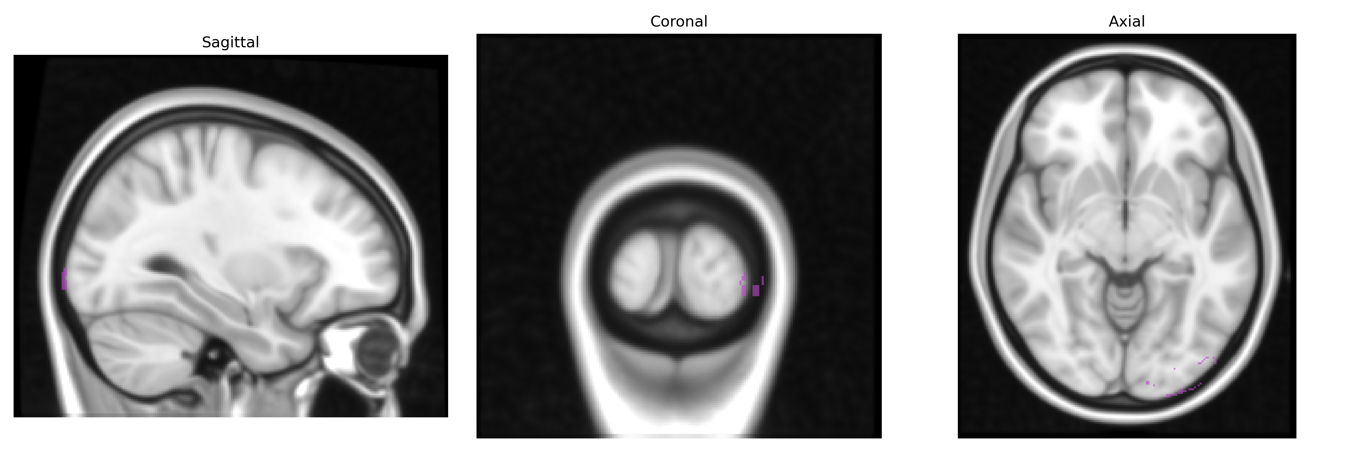
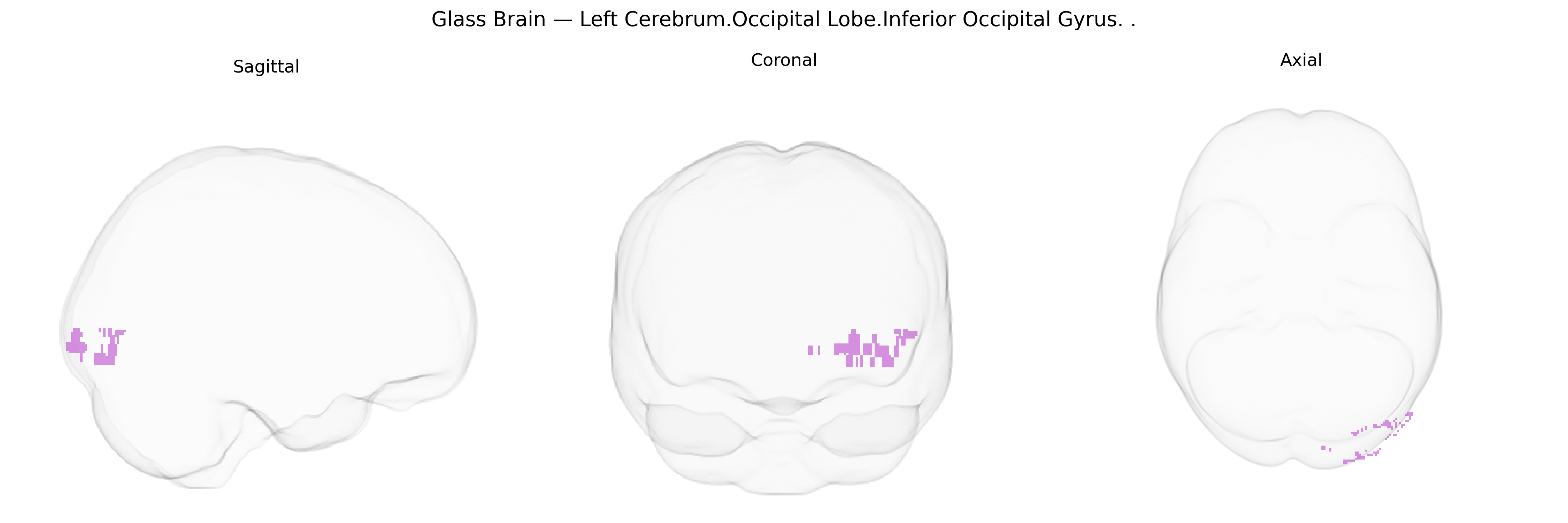
The bilateral Left Cerebrum.Occipital Lobe.Inferior Occipital Gyrus corresponds to a ventral occipital cortical region located on the inferior surface of the occipital lobe, bordering the temporal lobe and extending toward the occipitotemporal junction. It is primarily involved in early and intermediate stages of visual processing, including the analysis of shape, contour, and complex visual patterns, and contributes to object and face recognition pathways that project anteriorly into the ventral visual stream. Cytoarchitectonically, this region overlaps with areas such as Brodmann areas 18 and 19 and forms part of the occipitotemporal cortex that interfaces with higher-order visual regions in the fusiform and lateral occipital cortices. There is no direct link for the Inferior Occipital Gyrus; a closely related structure in the same region is the Occipital lobe.
The bilateral left cerebrum occipital lobe inferior occipital gyrus (IOG), a key ventral visual stream region implicated in object and face perception, shows convergent genetic associations from imaging–genetics and GWAS-based studies, although often at the level of broader occipital or ventral occipitotemporal cortex rather than Talairach-defined IOG alone. Large-scale brain MRI GWAS (e.g., ENIGMA, UK Biobank) have identified SNPs and loci influencing occipital cortical thickness, surface area, and volume near genes involved in neurodevelopment, synaptic function, and axon guidance (such as variants near HMGA2, IGF1, and neurodevelopmental genes affecting global and regional cortical morphology) that extend to inferior occipital/ventral visual areas. Polygenic risk for schizophrenia, major depressive disorder, and bipolar disorder has been associated with structural and functional alterations in inferior and lateral occipital regions, including reduced cortical thickness or altered activation during visual and face-processing tasks, suggesting shared genetic vulnerability affecting visual association cortex. Autism spectrum disorder genetic risk (e.g., rare CNVs and common polygenic burden) has also been linked to atypical activation and connectivity of inferior occipital and fusiform regions during social and face perception, implicating genes affecting synaptic plasticity and excitatory–inhibitory balance. In addition, occipital lobe epilepsies, sometimes associated with inherited channelopathies or receptor mutations (e.g., in genes encoding ion channels and GABAergic components), can involve the inferior occipital cortex, and migraine GWAS loci influencing visual cortex excitability may indirectly implicate this region. Overall, genetic influences on neurodevelopmental patterning, cortical arealization, and synaptic signaling appear to contribute to structural and functional variability in the inferior occipital gyrus, mediating susceptibility to psychiatric, neurodevelopmental, and visual processing–related traits, although precise, IOG-specific loci remain incompletely resolved and are typically inferred from broader occipital or ventral visual cortex analyses.
Overview generated by GPT-4o (2026).
Region ID: 251
Hemisphere: bilateral
Atlas: Talairach labels 1mm
Full Quality Version: Download MP4
Full Quality Version: Download MP4
Wali Sidiqyar*, Gaurav Rudravaram*, Elyssa M. McMaster, Trent M. Schwartz, Adam M. Saunders, Kurt G. Schilling, Bennett A. Landman "Introducing SPINS: A Shared Public Visualization Library of Neuroanatomical Structures." Medical Imaging with Deep Learning- short paper
This resource is licensed under CC0 1.0 Universal (Public Domain).