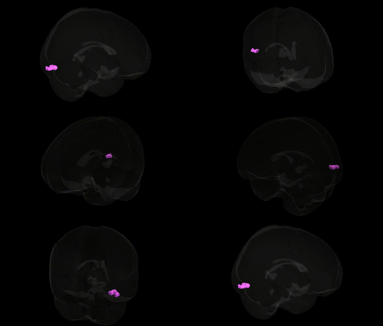
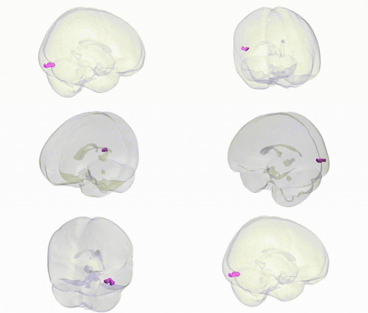
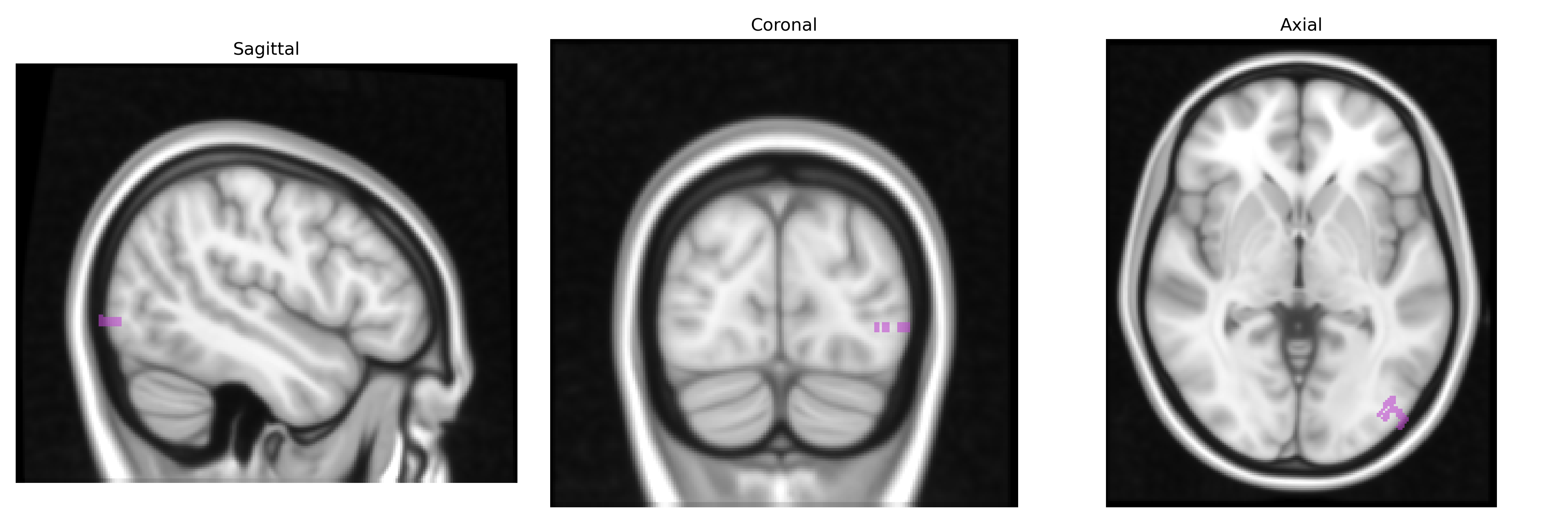
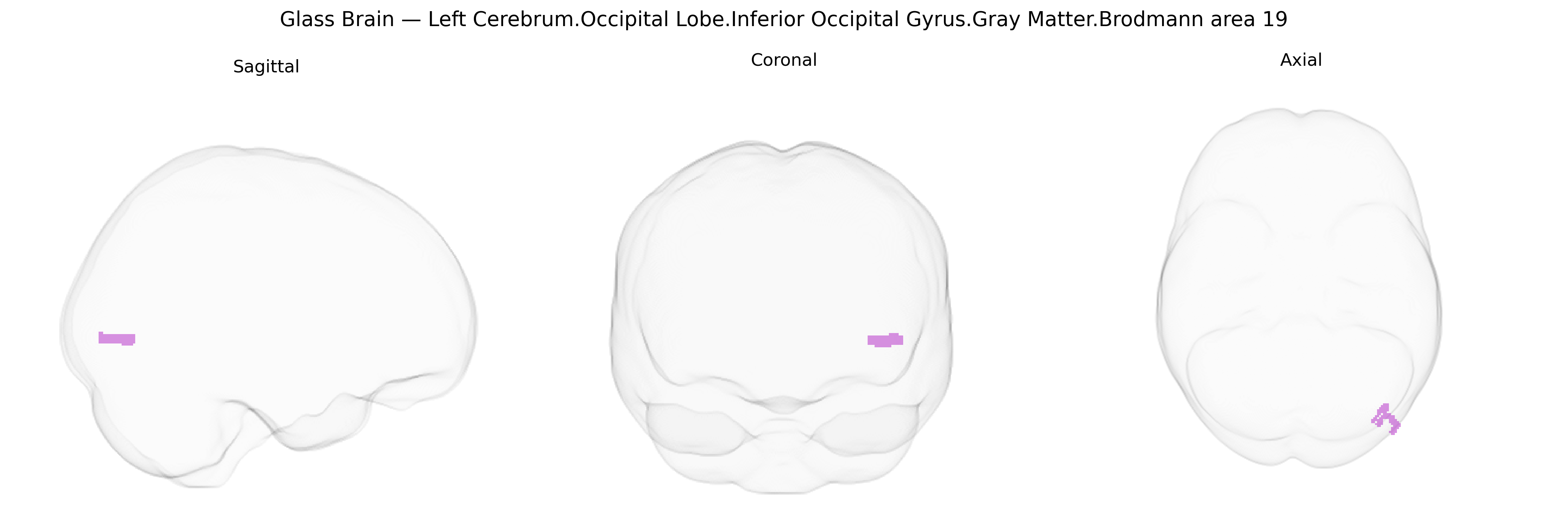
The bilateral Left Cerebrum.Occipital Lobe.Inferior Occipital Gyrus.Gray Matter.Brodmann area 19 corresponds to a portion of the associative visual cortex (BA19) located in the inferior aspect of the occipital lobe, bordering Brodmann areas 17 and 18 and extending toward occipitotemporal regions. This area participates in higher-order visual processing, including integration of complex visual features, object form, and aspects of motion and color, serving as a key node linking primary visual inputs to downstream temporal and parietal associative networks. Neuronal populations here exhibit columnar and laminar organization typical of neocortex and receive rich corticocortical inputs from primary and secondary visual cortices, as well as feedback projections from temporal and parietal association areas, supporting roles in visual recognition and visual–spatial analysis. No direct Wikipedia article exists for this exact composite region; a related structure is Brodmann area 19.
Genetic associations involving the inferior occipital gyrus gray matter in Brodmann area 19, a higher-order visual region, largely emerge from imaging-genetics and GWAS of occipital or visual cortex structure and function rather than region-specific candidate-gene studies. Common variants in genes regulating neurodevelopment and synaptic function—such as BDNF, NRG1, and glutamatergic or GABAergic pathway genes—have been linked to occipital cortical thickness, gray matter volume, or visual processing endophenotypes that include BA19, while polygenic scores for general cognitive ability and educational attainment show correlations with occipital gray matter measures in large cohorts (e.g., UK Biobank). Occipital and specifically BA18/19 regions appear in GWAS-based parcellation studies of cortical surface area and thickness, implicating loci near developmental transcription factors (e.g., PAX6, EMX2, LHX2) and axon-guidance genes, although these findings are typically reported at the level of “visual association cortex” rather than the inferior occipital gyrus alone. In neuropsychiatric GWAS, BA19 involvement is observed as an intermediate phenotype: risk variants for schizophrenia, autism spectrum disorder, and major depressive disorder are associated with altered occipital gray matter, functional connectivity, and visual processing anomalies, with imaging-genetics work implicating genes such as DISC1, CACNA1C, and complement-related loci in structural or functional differences encompassing BA19. In neurodegenerative and neuro-ophthalmologic contexts, APOE ε4 and other Alzheimer’s disease risk variants have been associated with posterior cortical atrophy patterns that include BA19, while GWAS of age-related macular degeneration and visual acuity highlight overlapping visual network changes, though direct gene–region specificity for the bilateral inferior occipital BA19 remains limited, with most evidence arising from broader visual association cortex phenotypes.
Overview generated by GPT-4o (2026).
Region ID: 414
Hemisphere: bilateral
Atlas: Talairach labels 1mm
Full Quality Version: Download MP4
Full Quality Version: Download MP4
Wali Sidiqyar*, Gaurav Rudravaram*, Elyssa M. McMaster, Trent M. Schwartz, Adam M. Saunders, Kurt G. Schilling, Bennett A. Landman "Introducing SPINS: A Shared Public Visualization Library of Neuroanatomical Structures." Medical Imaging with Deep Learning- short paper
This resource is licensed under CC0 1.0 Universal (Public Domain).