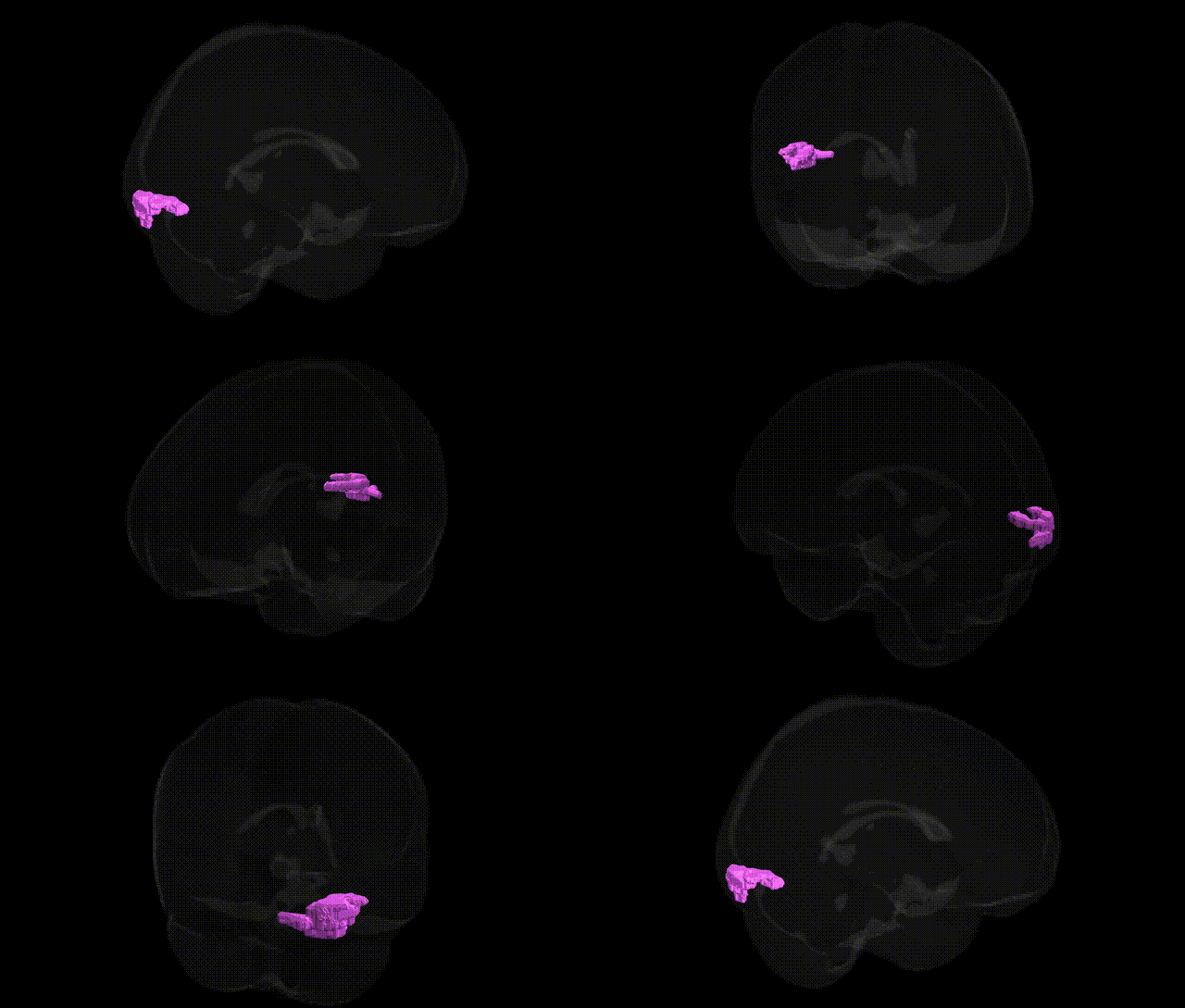
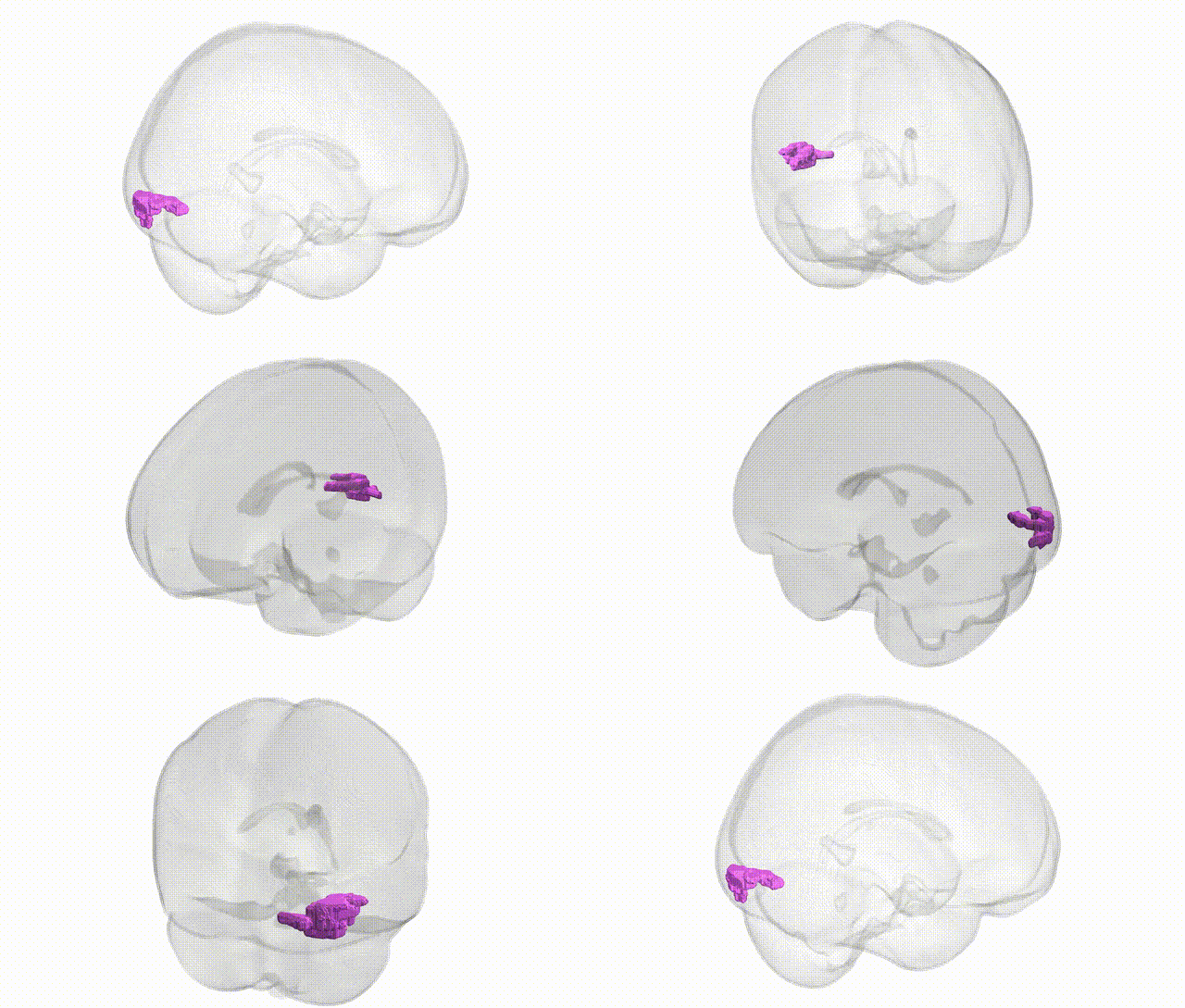
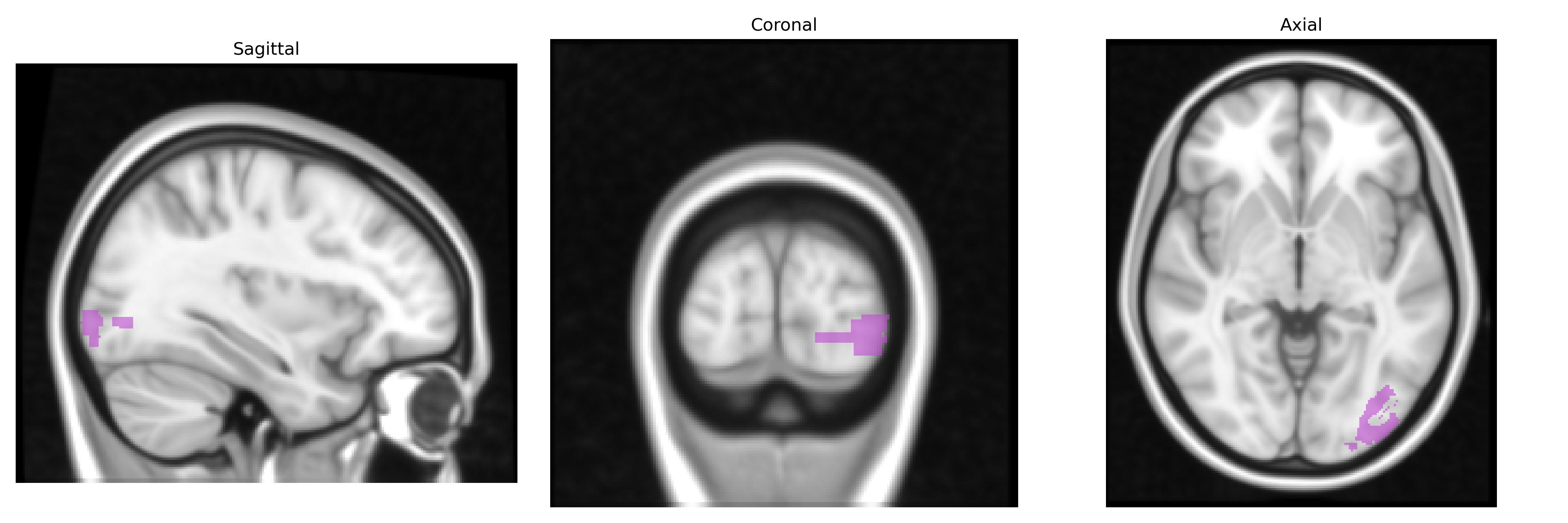
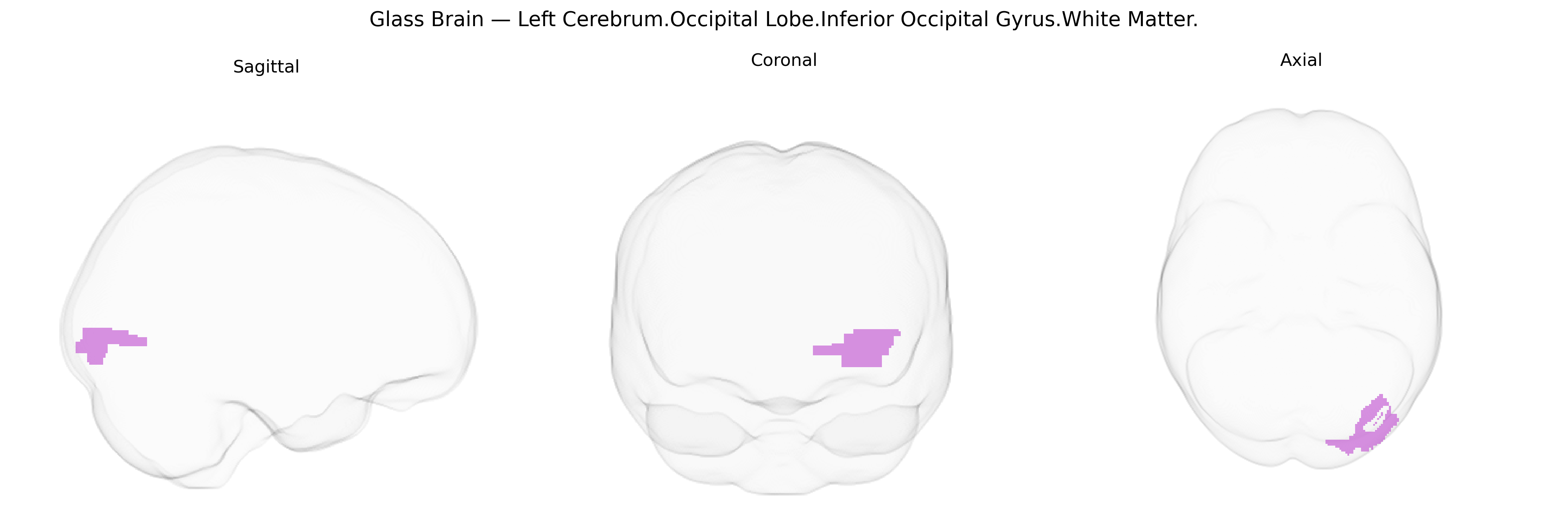
The bilateral Left Cerebrum.Occipital Lobe.Inferior Occipital Gyrus.White Matter corresponds to the deep myelinated fiber pathways underlying the inferior occipital gyrus in the left occipital lobe, as defined in the Talairach 1 mm atlas. This region contains association and projection fibers that interconnect primary and secondary visual cortices with temporal and parietal regions, contributing to the transmission and integration of visual information involved in object recognition and higher-order visual processing. It includes portions of major occipital white matter bundles, such as segments of the inferior longitudinal fasciculus and occipito-temporal association fibers, which form part of the ventral visual (“what”) pathway. There is no direct Wikipedia article for this specific white matter subdivision; a related structure is the Occipital lobe.
The bilateral left cerebrum occipital lobe inferior occipital gyrus white matter, as defined in the Talairach 1 mm atlas, lies within the broader occipital and visual association pathways for which genetic associations are typically reported at coarser regional or network levels rather than this exact tract; nonetheless, several large-scale imaging–genetics and GWAS efforts implicate genes and loci affecting occipital white matter microstructure, visual pathways, and related neuropsychiatric traits. Diffusion MRI GWAS (for example, UK Biobank–based studies) have identified common variants in genes involved in myelination, axon guidance, and oligodendrocyte function (such as NRG1/ERBB signaling components, MAG, and other glial-related genes) associated with fractional anisotropy and mean diffusivity in posterior white matter, including inferior occipital and occipito-temporal bundles. Occipital white matter integrity has been genetically linked, through polygenic risk and imaging endophenotype analyses, to schizophrenia, bipolar disorder, and major depressive disorder, where risk loci in synaptic and neurodevelopmental genes (e.g., CACNA1C, ZNF804A, MIR137-related networks) show associations with reduced occipital and visual pathway connectivity. Visual-processing and reading-related disorders, such as developmental dyslexia, have also been tied to genetic variants affecting posterior white matter tracts (including occipital-temporal pathways), with implicated genes like DCDC2, KIAA0319, and ROBO1 influencing axonal migration and cortical connectivity. In Alzheimer’s disease and other neurodegenerative conditions, GWAS loci such as APOE and CLU have been associated with widespread white matter changes involving posterior tracts, including occipital regions, as shown in imaging–genetic analyses linking these variants to altered diffusion metrics and posterior network disconnection. Overall, while direct GWAS hits specifically annotated to “inferior occipital gyrus white matter” are limited, convergent genetic evidence from white matter microstructure, visual pathway integrity, and neuropsychiatric and neurodevelopmental disorders supports a role for genes governing myelination, axonal development, and synaptic plasticity in shaping the structure and vulnerability of this posterior occipital white matter region.
Overview generated by GPT-4o (2026).
Region ID: 255
Hemisphere: bilateral
Atlas: Talairach labels 1mm
Full Quality Version: Download MP4
Full Quality Version: Download MP4
Wali Sidiqyar*, Gaurav Rudravaram*, Elyssa M. McMaster, Trent M. Schwartz, Adam M. Saunders, Kurt G. Schilling, Bennett A. Landman "Introducing SPINS: A Shared Public Visualization Library of Neuroanatomical Structures." Medical Imaging with Deep Learning- short paper
This resource is licensed under CC0 1.0 Universal (Public Domain).