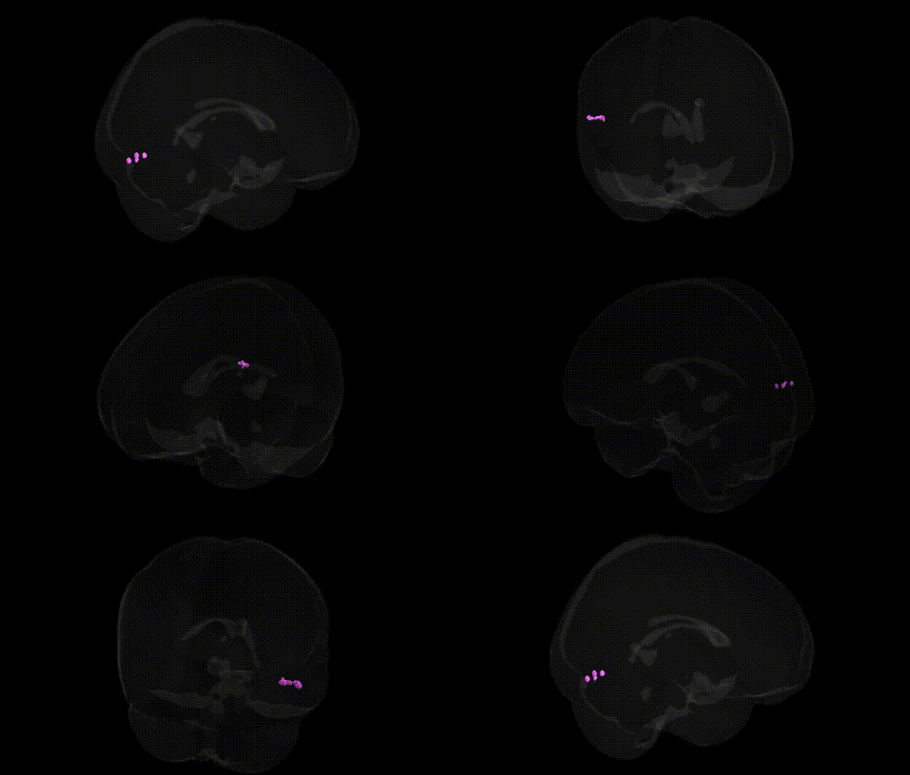
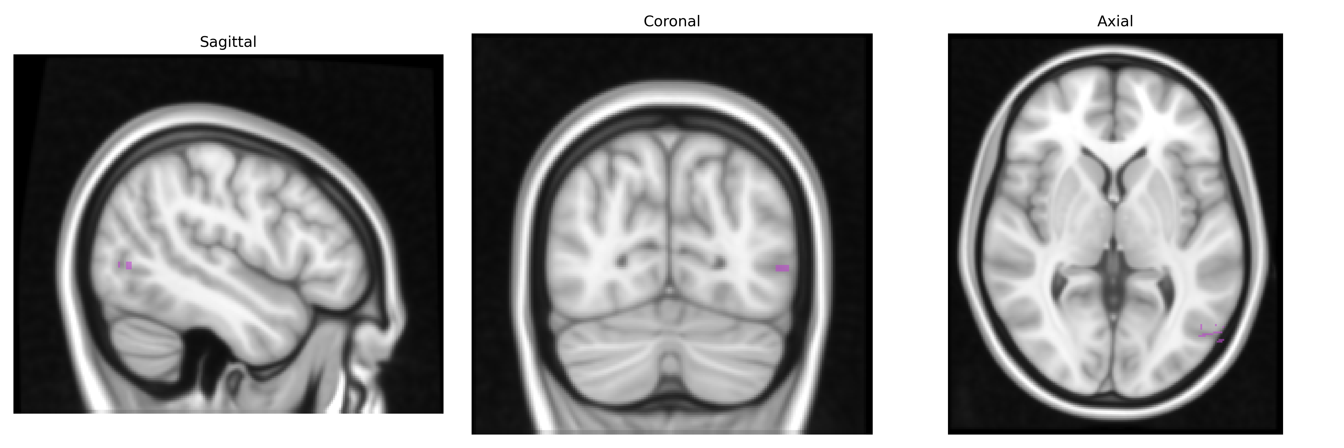
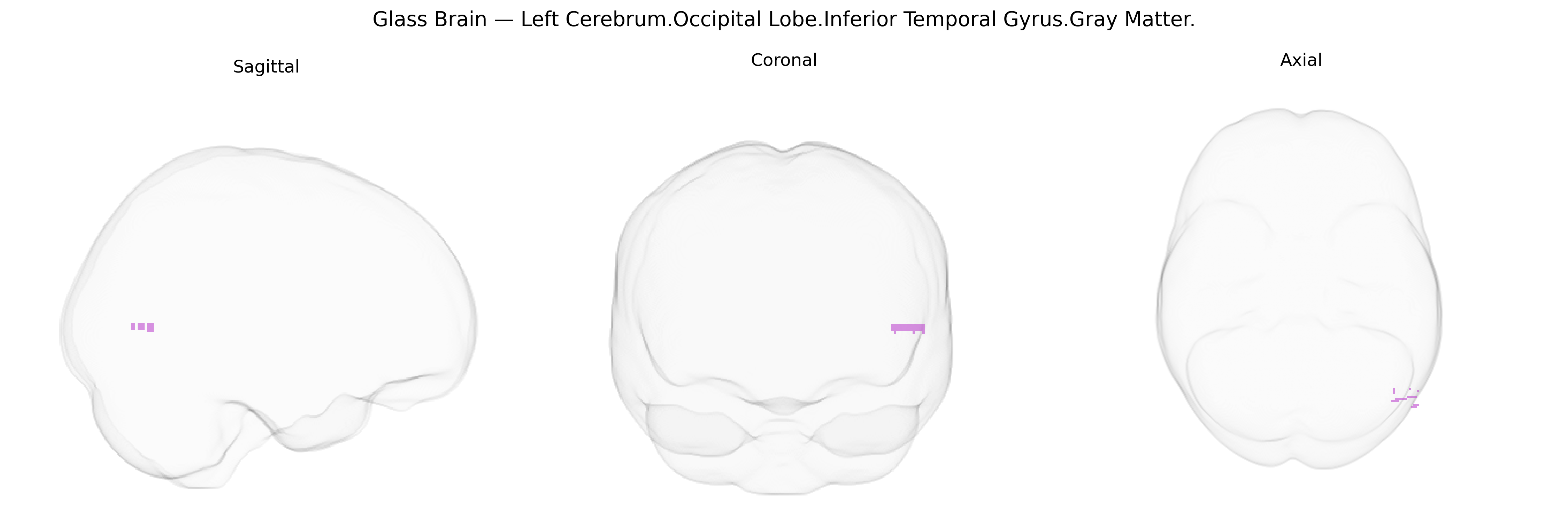
The bilateral Left Cerebrum.Occipital Lobe.Inferior Temporal Gyrus.Gray Matter corresponds to gray matter within the inferior temporal gyrus situated along the ventral surface of the occipital–temporal junction in the left hemisphere, as defined in the Talairach 1 mm atlas. This region is part of the ventral visual processing stream and is implicated in complex object, shape, and face recognition, as well as high-level visual association processes that link visual input with stored semantic information. It receives convergent input from earlier visual areas in the occipital lobe and projects to more anterior temporal and limbic structures, supporting integration of visual features into coherent percepts and contributing to category-specific processing (e.g., tools, animals, faces). Lesions involving this gray matter can lead to higher-order visual deficits such as visual agnosia or category-specific recognition impairments. There is no direct link for this exact Talairach label; a closely related structure is the Inferior temporal gyrus.
The inferior temporal gyrus (ITG) of the occipital/temporal junction, including bilateral gray matter regions corresponding to “Left Cerebrum.Occipital Lobe.Inferior Temporal Gyrus.Gray Matter” in Talairach-based atlases, is a key high-level visual and object-recognition area whose structure and function show substantial heritability and multiple genetic associations in imaging-genetics and GWAS work; large consortia such as ENIGMA and UK Biobank have reported common variants in genes involved in neurodevelopment, synaptic function, and cortical patterning (for example in or near MIR137, NPTN, POU3F2, and other neuronal and chromatin-regulation loci) associated with cortical thickness, surface area, and volume in ventral temporal/occipital regions encompassing the ITG, while polygenic risk scores for schizophrenia, major depressive disorder, autism spectrum disorder, and bipolar disorder have been linked to gray-matter alterations and cortical thinning in adjacent inferior temporal/ventral visual areas. Visual word-form and object-processing regions in the left ventral temporal/occipital cortex show structural and functional changes associated with risk or carrier status for neurodevelopmental and neurodegenerative disorders, including dyslexia-related and Alzheimer’s disease–related genetic risk (e.g., APOE ε4), and GWAS of cognitive traits such as intelligence, educational attainment, and face/object recognition performance have identified variants whose effects are partly mediated by morphology and connectivity of inferior temporal and fusiform cortices. Overall, genetic associations for this specific atlas-defined region are typically reported at the level of broader inferior temporal or ventral visual regions rather than the exact Talairach label, but converge on genes and polygenic profiles that influence cortical development, synaptic plasticity, and susceptibility to psychiatric and cognitive phenotypes manifesting in altered ITG structure and function.
Overview generated by GPT-4o (2026).
Region ID: 548
Hemisphere: bilateral
Atlas: Talairach labels 1mm
Full Quality Version: Download MP4
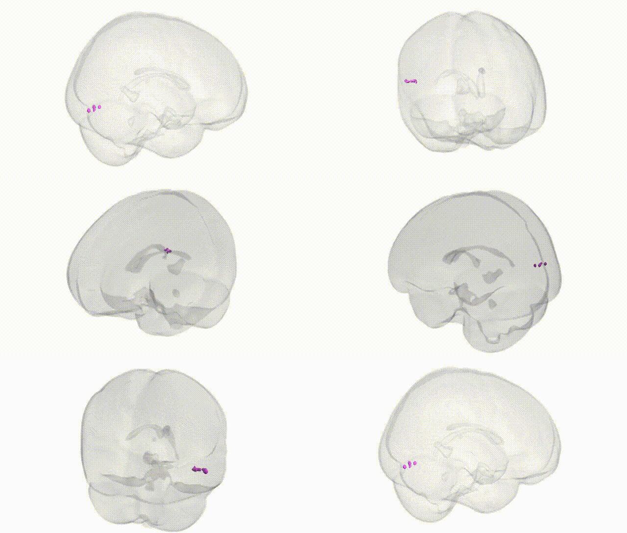
Full Quality Version: Download MP4
Wali Sidiqyar*, Gaurav Rudravaram*, Elyssa M. McMaster, Trent M. Schwartz, Adam M. Saunders, Kurt G. Schilling, Bennett A. Landman "Introducing SPINS: A Shared Public Visualization Library of Neuroanatomical Structures." Medical Imaging with Deep Learning- short paper
This resource is licensed under CC0 1.0 Universal (Public Domain).