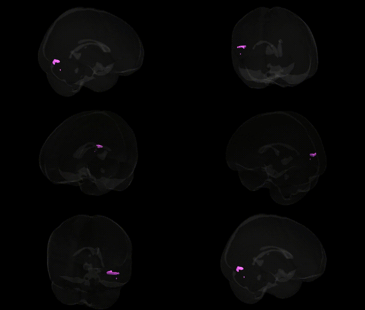
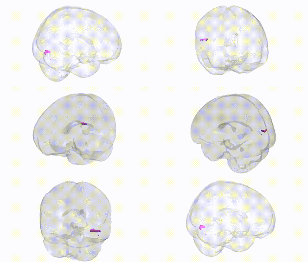
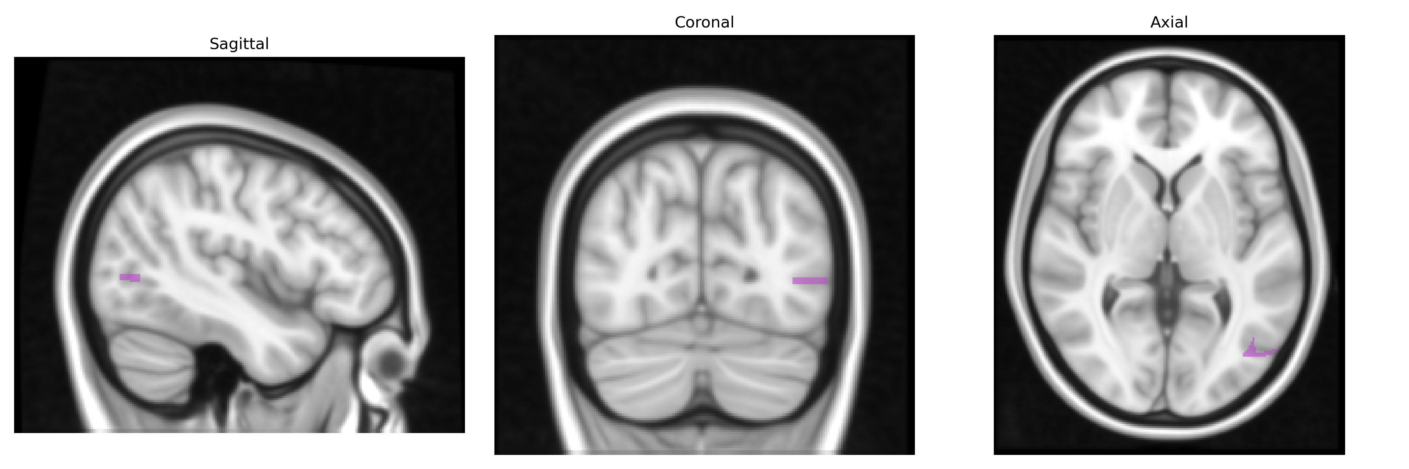
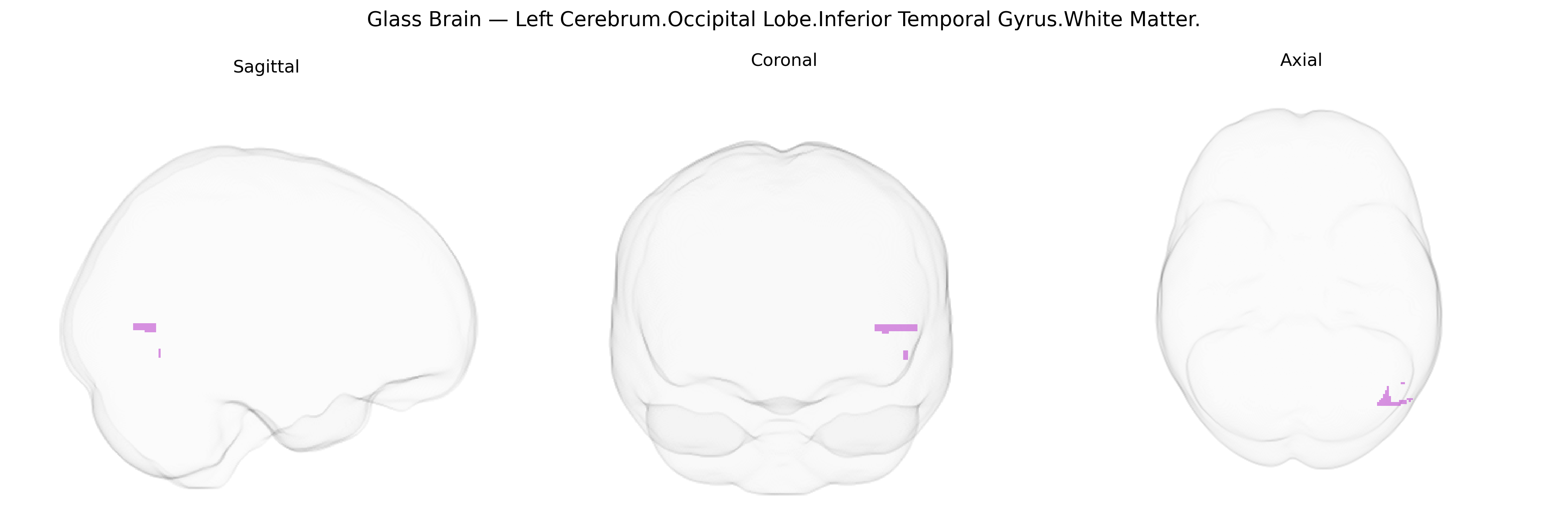
The bilateral Left Cerebrum.Occipital Lobe.Inferior Temporal Gyrus.White Matter corresponds to deep intrahemispheric fiber pathways coursing beneath the inferior temporal gyrus at the occipital–temporal junction, supporting communication between visual association cortices, ventral temporal regions, and more anterior temporal and frontal areas. These white matter tracts participate in the ventral visual (“what”) pathway, relaying high-level object, face, and word-form information from occipital visual areas toward temporal and limbic structures involved in recognition and memory. They include contributions from long association bundles such as the inferior longitudinal fasciculus and related fibers that link occipital cortex with anterior temporal regions, enabling integration of complex visual input with semantic processing and episodic memory networks. There is no direct link for this exact Talairach-defined region; a related structure is the Inferior Temporal Gyrus.
The bilateral left cerebrum occipital lobe inferior temporal gyrus white matter, as defined in the Talairach 1 mm atlas, lies within ventral visual and object-recognition pathways, and genetic associations involving this region generally derive from imaging genetics and GWAS of brain structure rather than from studies of the specific label. Large-scale GWAS of cortical thickness, surface area, and white-matter microstructure (e.g., ENIGMA and UK Biobank studies) have identified numerous loci (including variants near genes such as HMGA2, PAX6, DCC, and others implicated in neurodevelopment) that influence occipito-temporal morphology and fractional anisotropy in adjacent tracts (such as the inferior longitudinal fasciculus) supporting visual object and face processing. Alterations in white matter of the inferior temporal/occipital region have been associated, in genetically informed studies, with neurodevelopmental and neuropsychiatric conditions including autism spectrum disorder, schizophrenia, and dyslexia, where polygenic risk scores correlate with microstructural differences in ventral visual pathways. Rare variants and CNVs in genes affecting axon guidance, synaptic development, and myelination (e.g., ROBO/SLIT, CNTNAP2, and others) have been linked to disruptions of occipito-temporal connectivity that encompass this region, and GWAS on face recognition and reading-related traits have also implicated nearby occipito-temporal territories (such as the visual word form area) whose white-matter connections traverse the inferior temporal gyrus. However, current genetic literature typically reports associations at the level of broader occipital or temporal lobes and major white-matter tracts rather than specifically isolating the Talairach-defined “Left Cerebrum.Occipital Lobe.Inferior Temporal Gyrus.White Matter” parcel.
Overview generated by GPT-4o (2026).
Region ID: 326
Hemisphere: bilateral
Atlas: Talairach labels 1mm
Full Quality Version: Download MP4
Full Quality Version: Download MP4
Wali Sidiqyar*, Gaurav Rudravaram*, Elyssa M. McMaster, Trent M. Schwartz, Adam M. Saunders, Kurt G. Schilling, Bennett A. Landman "Introducing SPINS: A Shared Public Visualization Library of Neuroanatomical Structures." Medical Imaging with Deep Learning- short paper
This resource is licensed under CC0 1.0 Universal (Public Domain).