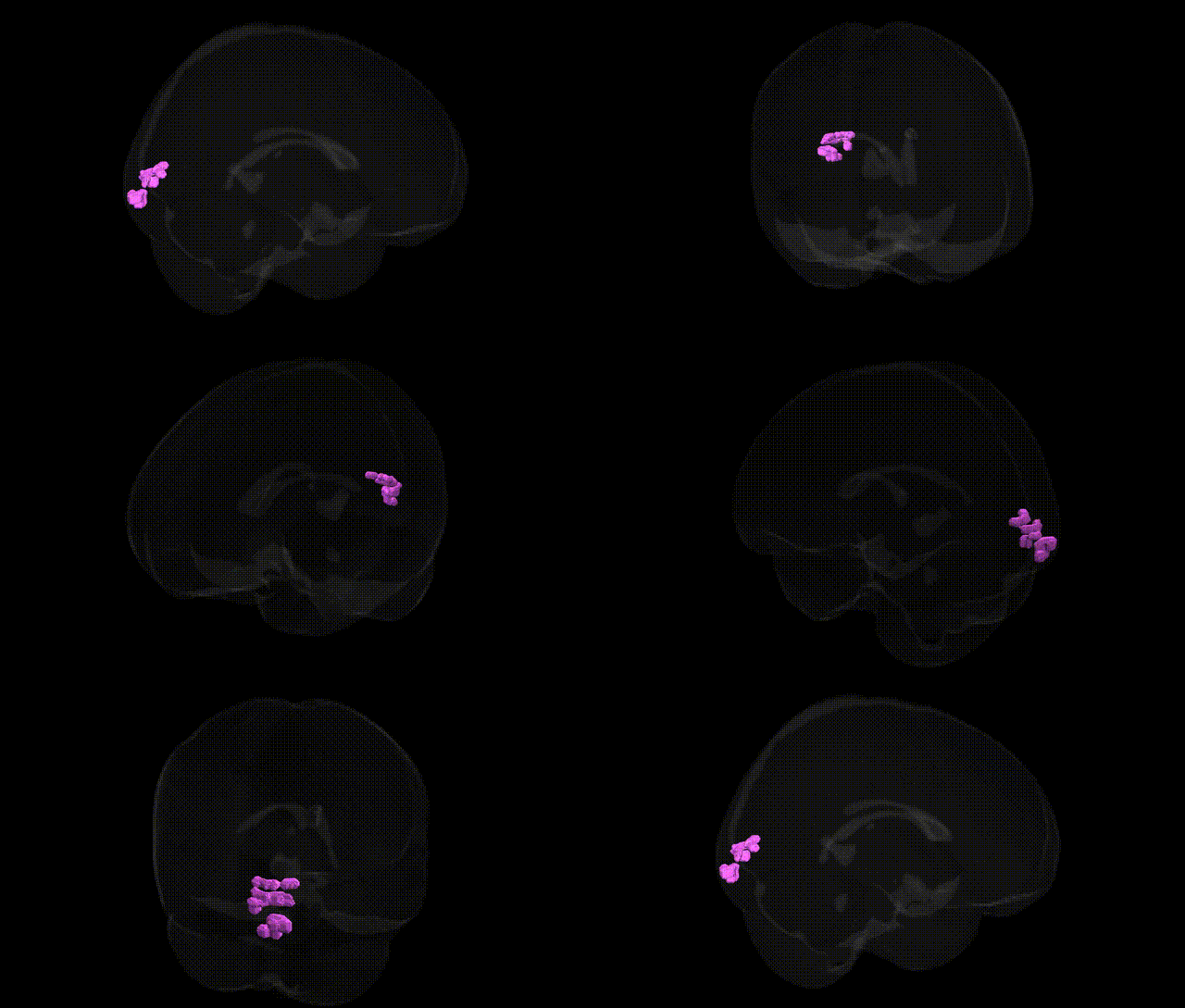
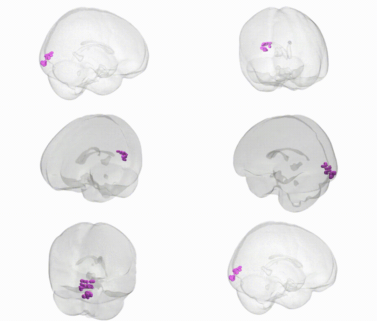
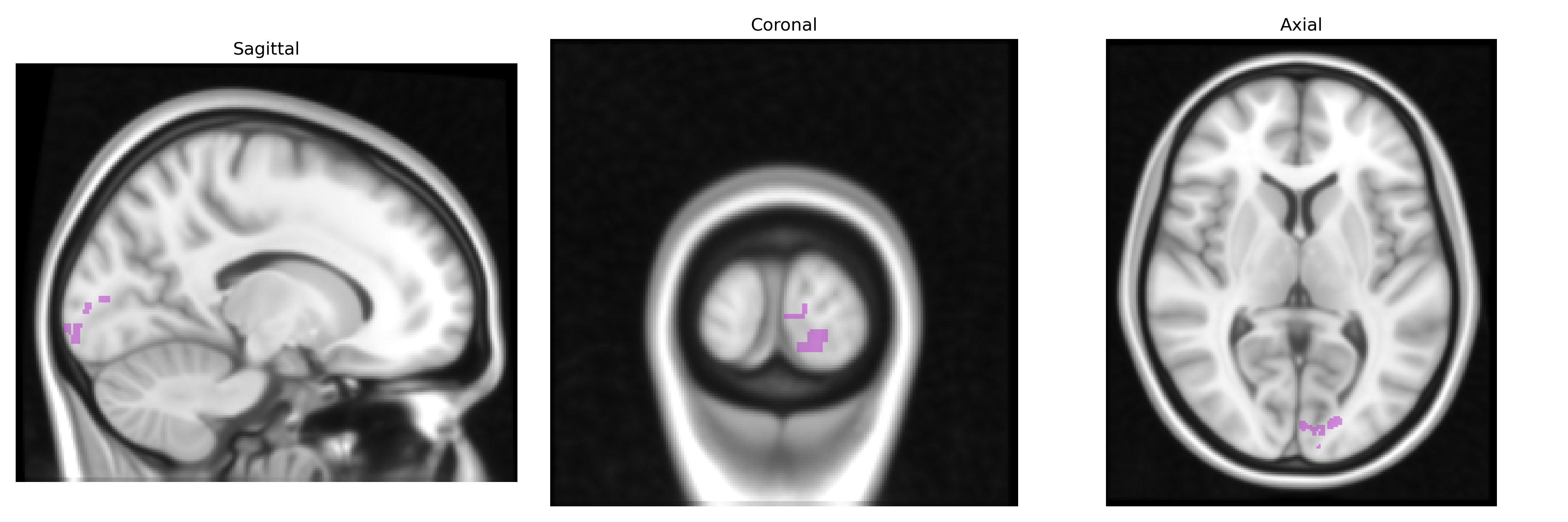
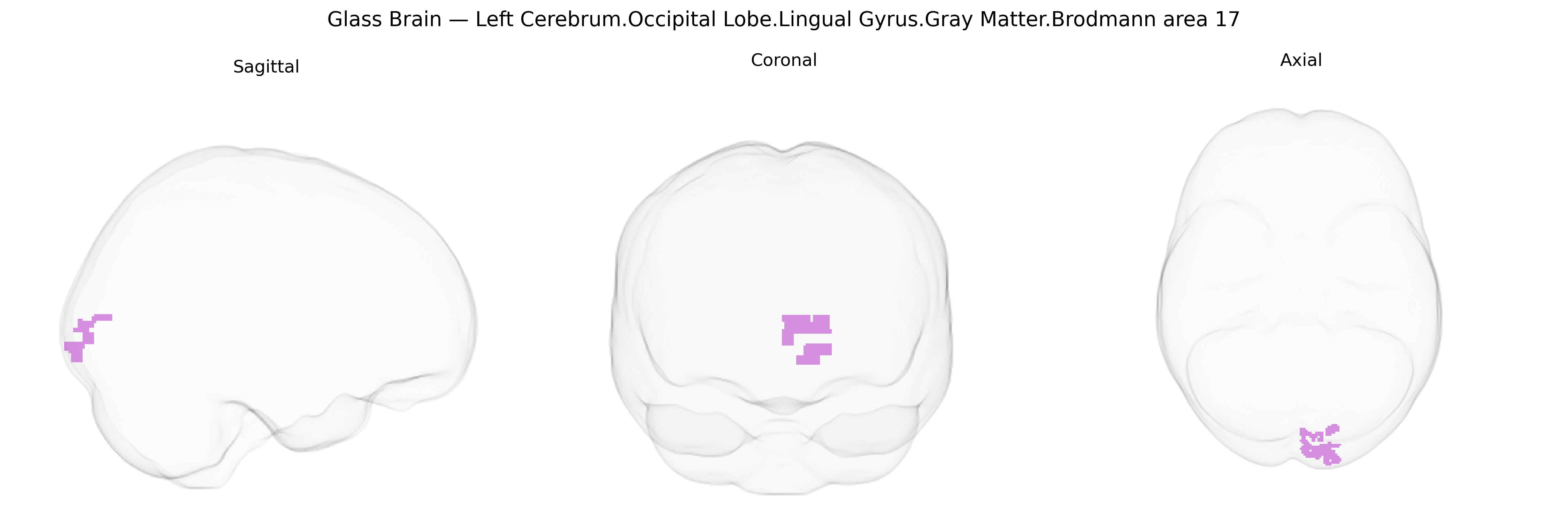
The bilateral Left Cerebrum.Occipital Lobe.Lingual Gyrus.Gray Matter.Brodmann area 17 corresponds to primary visual cortex (V1) located along the calcarine fissure within the lingual gyrus of the occipital lobe. This region represents the initial cortical stage of visual processing, receiving highly organized retinotopic input from the lateral geniculate nucleus of the thalamus. Neurons in Brodmann area 17 are specialized for detecting basic visual features such as orientation, spatial frequency, contrast, and motion direction, forming the foundation for higher-order visual analysis in adjacent extrastriate areas. The laminar organization and dense myelination of this cortex reflect its role as a major thalamorecipient zone, crucial for conscious visual perception and the construction of precise visual maps of the contralateral visual field. Brodmann area 17
The bilateral left occipital lingual gyrus gray matter within Brodmann area 17 (primary visual cortex) has been implicated in a range of genetically influenced traits, primarily through imaging-genetics and GWAS of cortical structure and visual, cognitive, and psychiatric phenotypes. Large-scale MRI GWAS consortia (e.g., ENIGMA, UK Biobank) have identified variants in genes involved in neurodevelopment, axon guidance, and synaptic function (such as HMGA2, MIR423, and loci near KIAA0586, DACT1, and other Wnt/SHH-pathway genes) associated with occipital and specifically lingual gyrus cortical thickness, surface area, and volume, although most findings map to broader occipital or visual cortex regions rather than BA17 alone. Polygenic risk for schizophrenia, bipolar disorder, and major depression has been linked to structural and functional alterations in the lingual gyrus/BA17, with several case–control and polygenic score studies showing that risk alleles for these disorders correlate with reduced gray matter volume or altered activation in this region. In neurodevelopmental and neuro-ophthalmologic conditions, rare variants and CNVs affecting genes critical for visual pathway development (e.g., PAX6, SOX2, OTX2, and other transcription factors) have been associated with anomalies of primary visual cortex that include the calcarine and lingual regions, contributing to cortical visual impairment and albinism-related misrouting of visual projections. GWAS of visual acuity, refractive error, and visual processing speed also implicate loci near genes involved in retinal and cortical visual pathway development, some of which show downstream effects on occipital cortical structure. Additionally, imaging-genetic studies of reading ability, dyslexia, and visual word recognition indicate that allelic variation in genes modulating neuronal migration and myelination (e.g., DCDC2, KIAA0319, and other candidate dyslexia genes) can influence function and microstructure in the left lingual and adjacent fusiform regions, though associations are typically distributed across a broader ventral occipitotemporal network. Overall, genetic findings for this specific Talairach-defined region converge on polygenic influences on early visual cortex development, occipital morphology, and susceptibility to psychiatric and neurodevelopmental disorders with visual and visuo-cognitive components, but no single gene or variant is uniquely or exclusively associated with the left BA17 lingual gyrus.
Overview generated by GPT-4o (2026).
Region ID: 242
Hemisphere: bilateral
Atlas: Talairach labels 1mm
Full Quality Version: Download MP4
Full Quality Version: Download MP4
Wali Sidiqyar*, Gaurav Rudravaram*, Elyssa M. McMaster, Trent M. Schwartz, Adam M. Saunders, Kurt G. Schilling, Bennett A. Landman "Introducing SPINS: A Shared Public Visualization Library of Neuroanatomical Structures." Medical Imaging with Deep Learning- short paper
This resource is licensed under CC0 1.0 Universal (Public Domain).