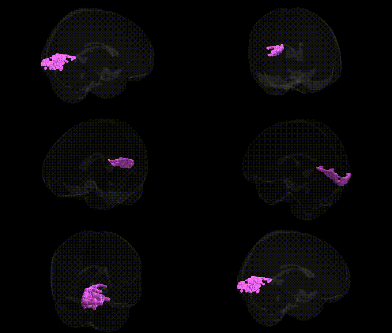
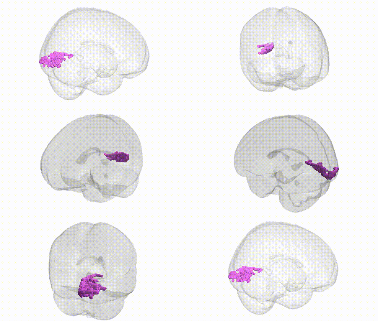
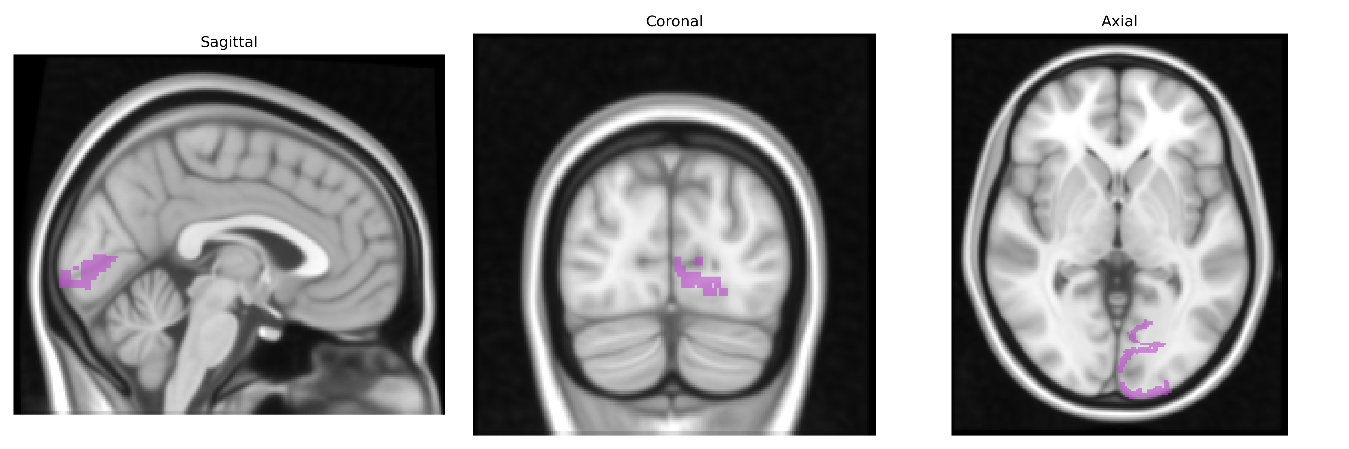
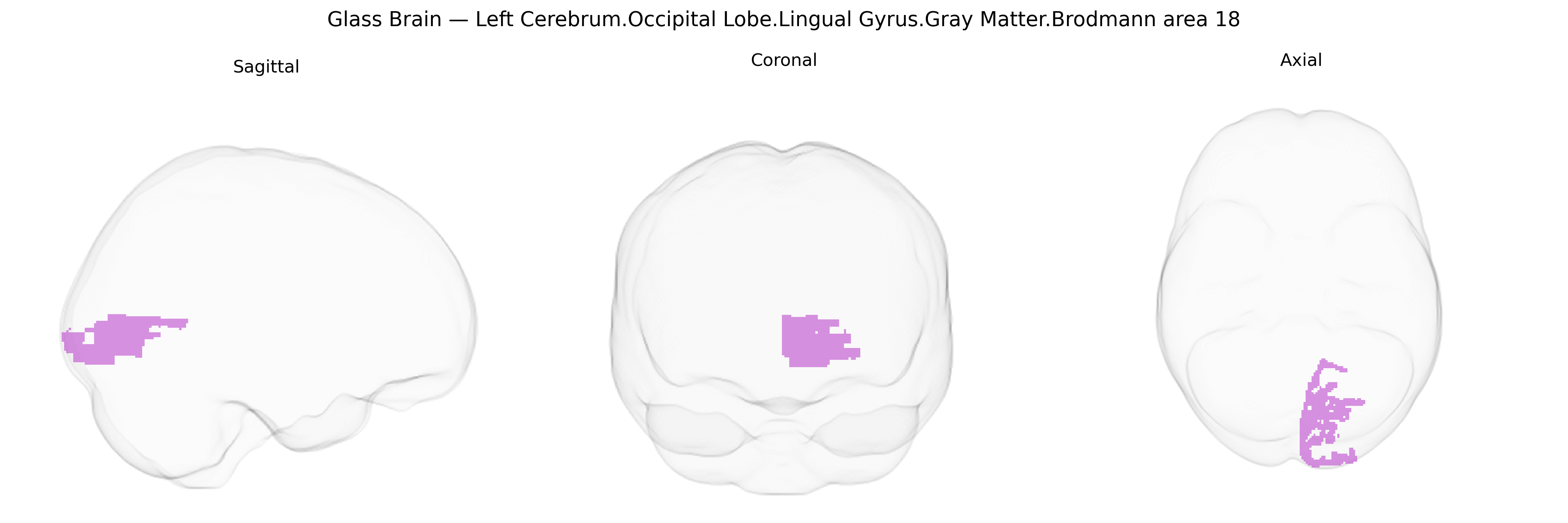
The bilateral Left Cerebrum.Occipital Lobe.Lingual Gyrus.Gray Matter.Brodmann area 18 corresponds to secondary visual cortex (V2) located in the lingual gyrus of the occipital lobe, just anterior to primary visual cortex (V1, BA17). This region is involved in early-stage visual processing, including analysis of orientation, spatial frequency, color, and more complex visual features that contribute to pattern and form recognition. Cytoarchitectonically, BA18 is characterized by a well-developed granular layer IV and distinct laminar differentiation relative to BA17, reflecting its role in integrating feedforward input from V1 with feedback and associative projections from higher visual areas. Functionally, activity in this region supports contour integration, binocular disparity processing, and the transformation of basic visual signals into more structured percepts that can be utilized by downstream dorsal and ventral visual pathways. There is no direct Wikipedia article for this specific lingual-gyrus-defined BA18 region; a related structure is Brodmann area 18.
The left lingual gyrus (Brodmann area 18) in the occipital lobe is a secondary visual association region implicated in genetic studies of visual processing, reading, and neuropsychiatric phenotypes. Twin and imaging–genetics studies show high heritability of gray matter volume and cortical thickness in this area, with polygenic influences distributed across many loci rather than a single major gene. GWAS of brain structure (e.g., ENIGMA, UK Biobank) have reported associations between variants near genes involved in neurodevelopment and synaptic function (such as those in Wnt signaling, axon guidance, and chromatin regulation pathways) and occipital/lingual gyrus morphology, though specific effects are typically small and regionally overlapping. Functionally, genetic risk for schizophrenia, major depressive disorder, bipolar disorder, and autism spectrum disorders has been linked to altered activation or structure of BA18/lingual gyrus in PRS and case–control imaging–genetics analyses, often in the context of visual perception, emotion processing, or hallucinations. Variants associated with dyslexia and reading-related traits have been connected to altered activation in left occipital and occipitotemporal regions that include or border the lingual gyrus, while migraine, visual aura, and occipital lobe epilepsy studies occasionally implicate this area through networks influenced by ion channel and neurotransmission genes (e.g., CACNA1A and related pathways), although links are generally network-level rather than region-specific. Overall, genetic findings support a polygenic, pleiotropic architecture in which many common variants shape the structure and function of left BA18/lingual gyrus as part of wider visual and cognitive networks rather than a discrete set of region-exclusive risk genes.
Overview generated by GPT-4o (2026).
Region ID: 247
Hemisphere: bilateral
Atlas: Talairach labels 1mm
Full Quality Version: Download MP4
Full Quality Version: Download MP4
Wali Sidiqyar*, Gaurav Rudravaram*, Elyssa M. McMaster, Trent M. Schwartz, Adam M. Saunders, Kurt G. Schilling, Bennett A. Landman "Introducing SPINS: A Shared Public Visualization Library of Neuroanatomical Structures." Medical Imaging with Deep Learning- short paper
This resource is licensed under CC0 1.0 Universal (Public Domain).