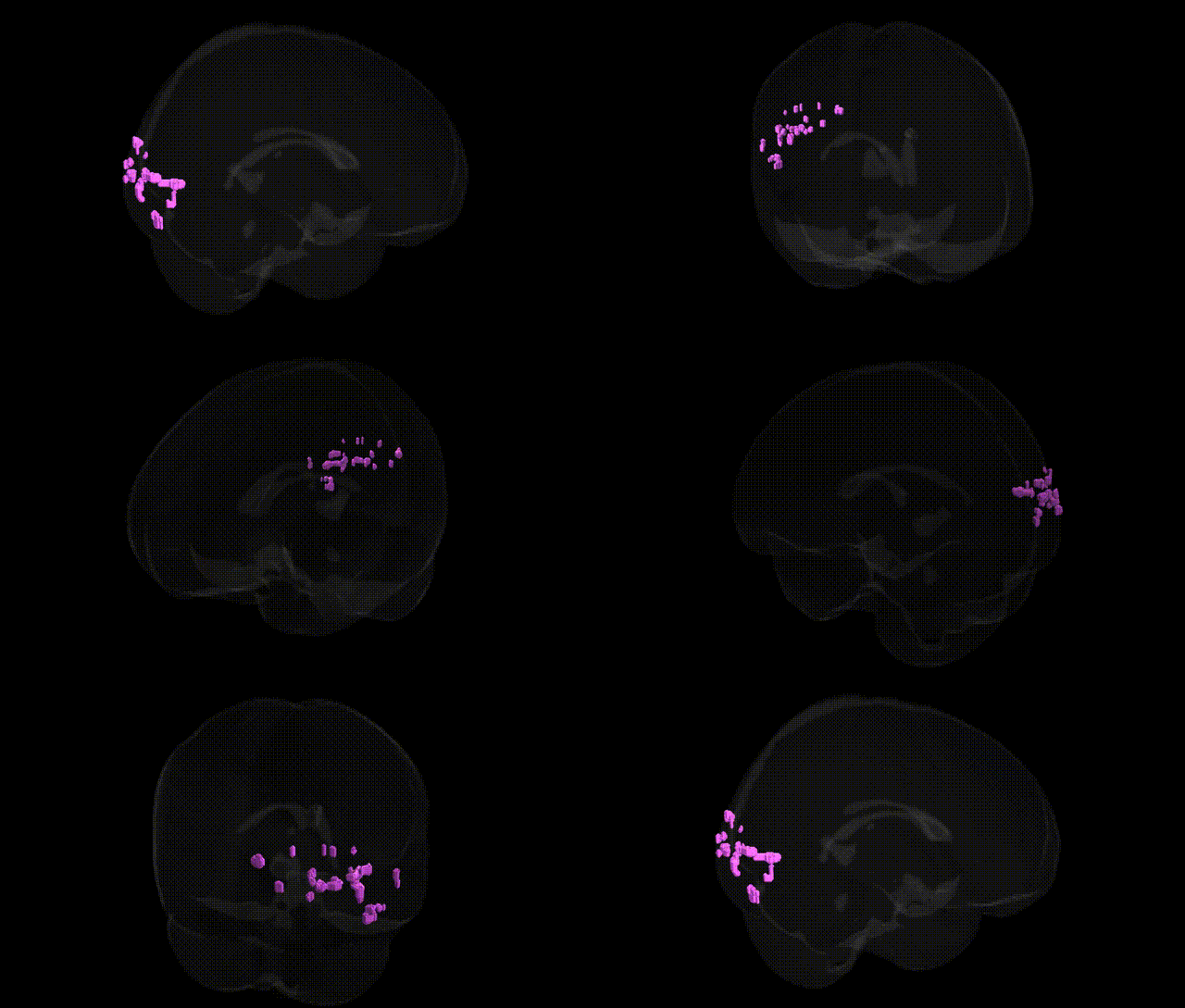
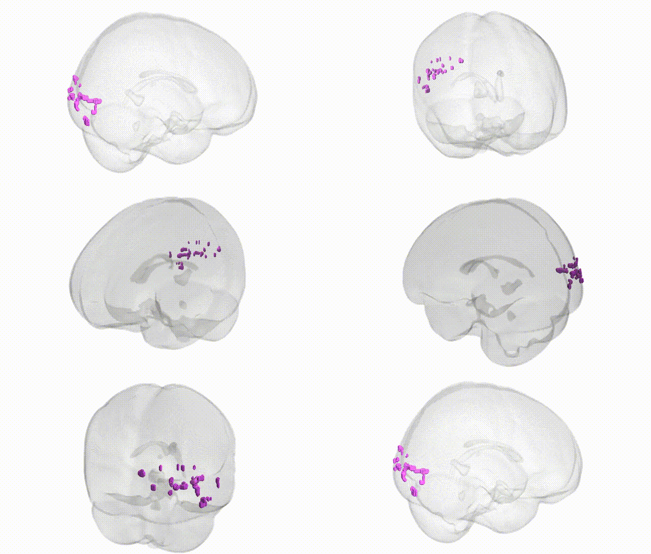
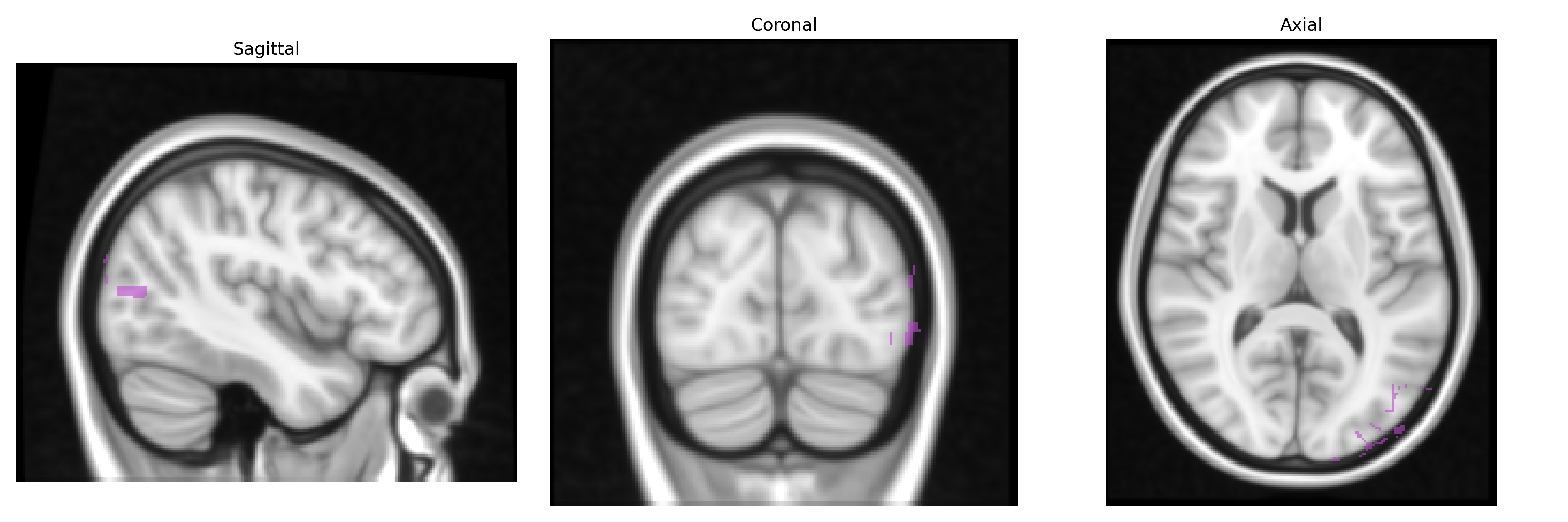
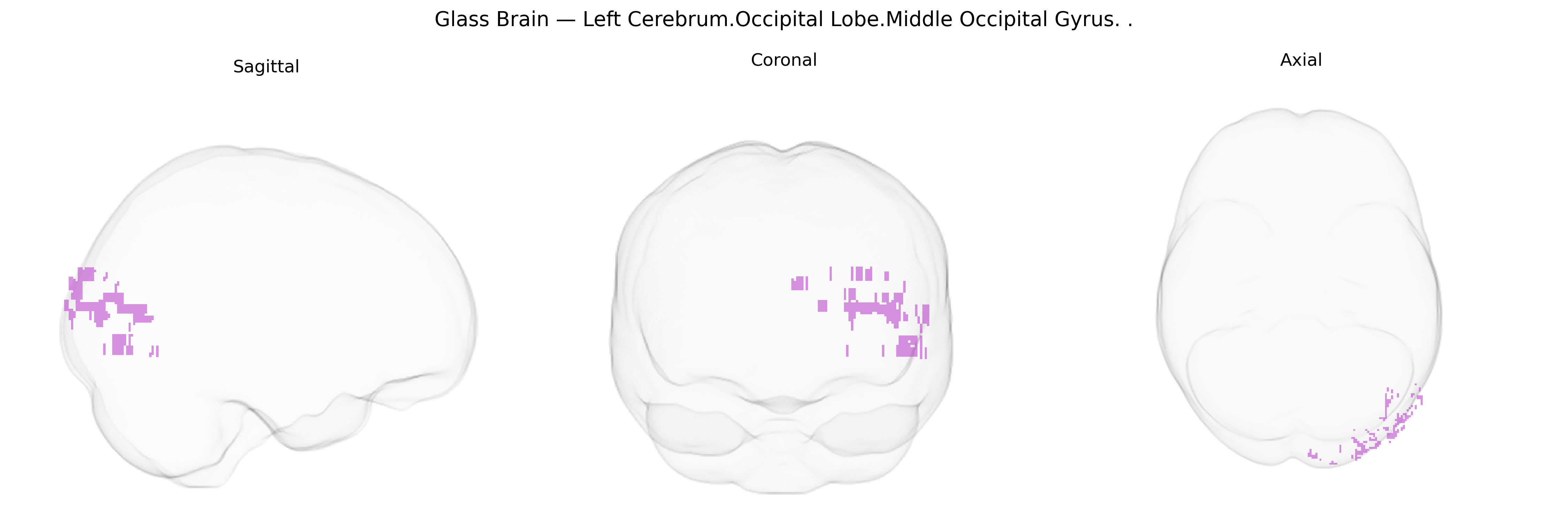
The bilateral Left Cerebrum.Occipital Lobe.Middle Occipital Gyrus corresponds to the middle portion of the lateral occipital cortex, situated between the superior and inferior occipital gyri in the occipital lobe, and is primarily involved in intermediate-level visual processing. This region contributes to the analysis of object form, motion, and spatial relationships, integrating inputs from primary visual areas and relaying processed information to higher-order visual and multisensory association cortices. Functionally, it participates in recognition of shapes and complex visual patterns and is implicated in tasks requiring visuospatial attention and object perception. There is no direct link; see the related structure Occipital lobe.
The bilateral left middle occipital gyrus (Talairach labels 1 mm Atlas: Left Cerebrum.Occipital Lobe.Middle Occipital Gyrus) is a core part of the lateral occipital visual cortex, and genetic associations involving this region largely emerge from imaging‑genetics and GWAS of brain structure, functional activation, and visual‐related traits rather than from region‑exclusive findings. Large MRI‑GWAS consortia (e.g., ENIGMA, UK Biobank) have identified common variants in neurodevelopmental and axon‑guidance genes (such as those near MAPT, HMGA2, and several loci enriched for synaptic and neuronal differentiation pathways) that associate with occipital cortical thickness or surface area, with effects that extend to the middle occipital gyrus among broader occipital measures. Functional imaging‑genetics studies show that variants influencing visual processing, face and object recognition, and reading (including loci near DCDC2, KIAA0319, and other dyslexia‑related genes) can modulate activation patterns in lateral occipital regions encompassing the middle occipital gyrus. This region’s structure and function have been implicated in neurodevelopmental and psychiatric conditions—such as schizophrenia, autism spectrum disorder, and attention‑deficit/hyperactivity disorder—through case–control and polygenic risk studies reporting altered occipital morphology or activation, often tied to polygenic load for these disorders rather than to single genes. Additional associations link occipital morphology or perfusion, including the middle occipital gyrus, with genetic risk for migraine (e.g., variants in TRPM8 and other pain/vascular‑related loci), visual hallucinations in Parkinson’s disease, and visual‑spatial or reading performance traits, although these results typically involve networks of occipital and parietal regions rather than the middle occipital gyrus in isolation.
Overview generated by GPT-4o (2026).
Region ID: 310
Hemisphere: bilateral
Atlas: Talairach labels 1mm
Full Quality Version: Download MP4
Full Quality Version: Download MP4
Wali Sidiqyar*, Gaurav Rudravaram*, Elyssa M. McMaster, Trent M. Schwartz, Adam M. Saunders, Kurt G. Schilling, Bennett A. Landman "Introducing SPINS: A Shared Public Visualization Library of Neuroanatomical Structures." Medical Imaging with Deep Learning- short paper
This resource is licensed under CC0 1.0 Universal (Public Domain).