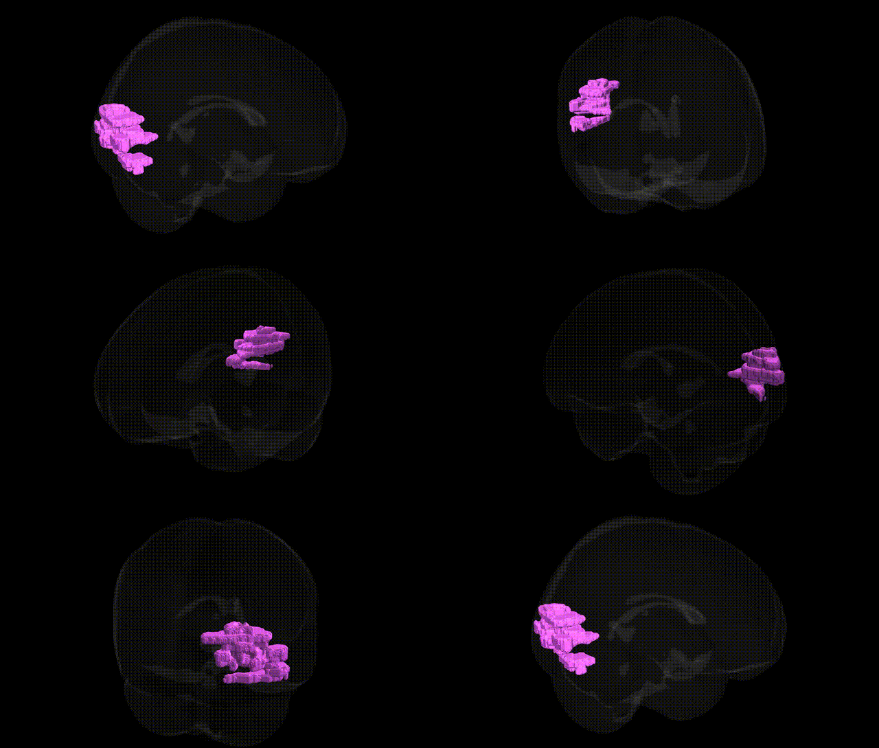
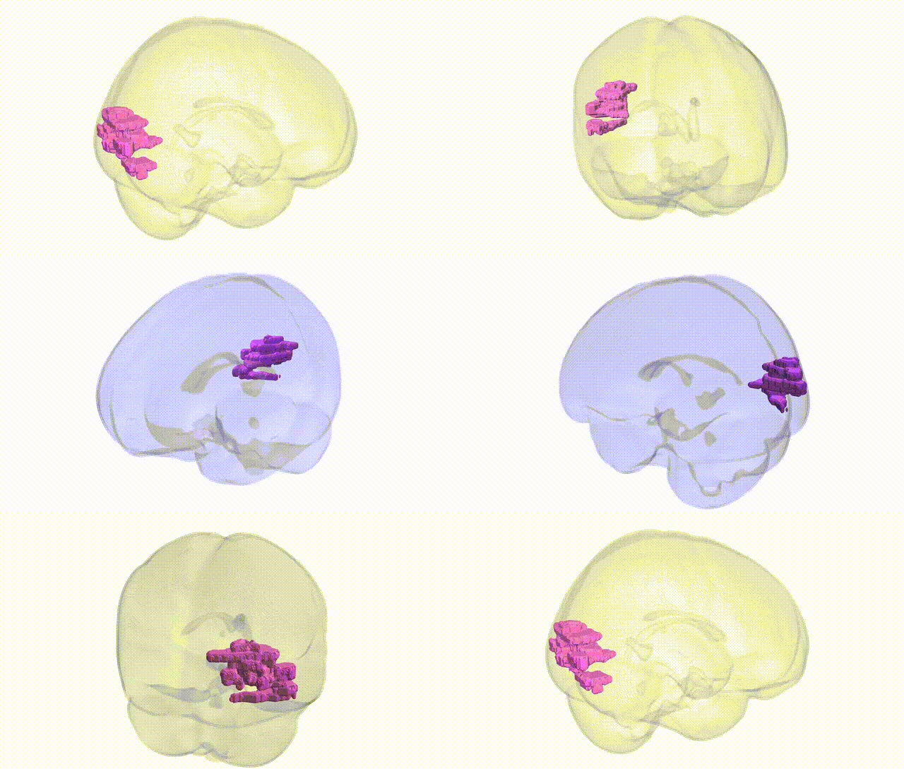
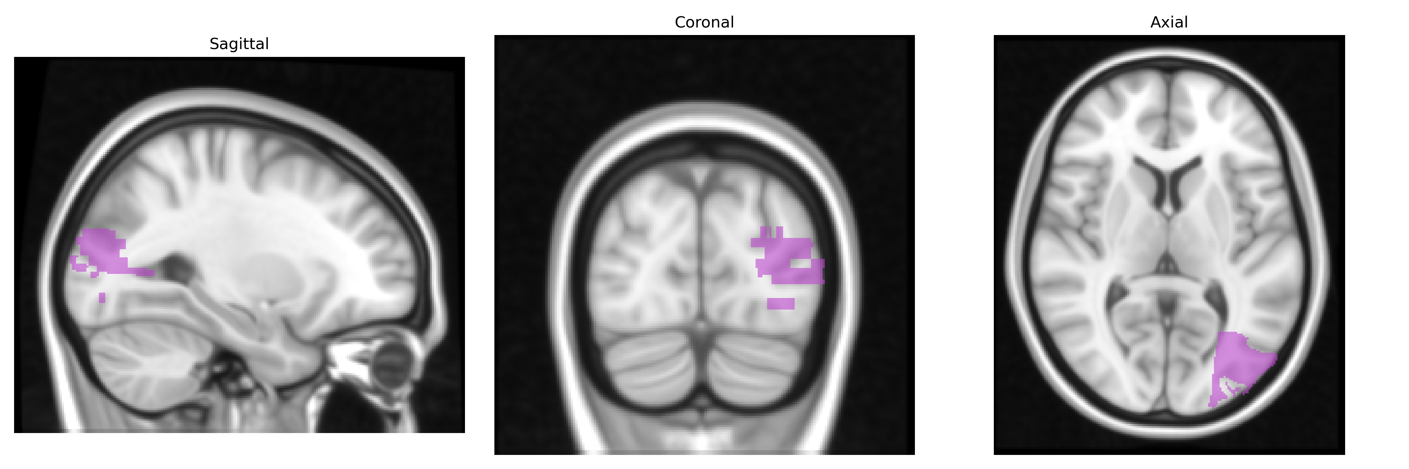
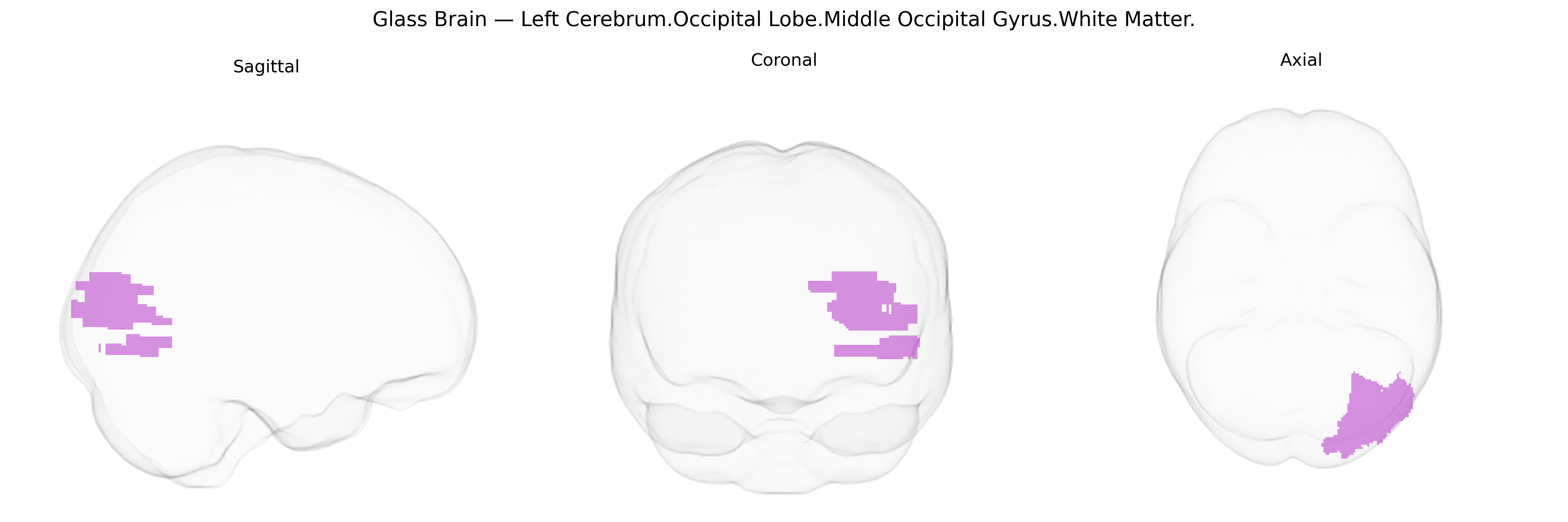
The bilateral Left Cerebrum.Occipital Lobe.Middle Occipital Gyrus.White Matter corresponds to the subcortical myelinated fiber pathways underlying the middle occipital gyrus in the occipital lobe, as defined in the Talairach 1 mm atlas. This white matter region carries visual information between primary and secondary visual areas and higher-order association cortices, contributing to the integration of motion, form, and spatial aspects of vision. It includes portions of long association tracts such as the inferior longitudinal fasciculus and possibly segments of the inferior fronto-occipital fasciculus, which link occipital visual regions with temporal and frontal areas for object recognition and visuospatial processing. There is no direct Wikipedia article for this specific white matter label; related information can be found under the broader Occipital lobe.
Genetic associations specific to the bilateral Left Cerebrum Occipital Lobe Middle Occipital Gyrus white matter, as defined in the Talairach 1 mm atlas, are not well resolved at the fine-grain regional level, but convergent evidence from imaging–genetics and GWAS implicates nearby occipital white-matter pathways and visual-association regions in several heritable traits and disorders. Twin and SNP-heritability studies show high heritability of occipital white-matter microstructure (e.g., fractional anisotropy in optic radiations and adjacent association fibers), with implicated genes often involving axon guidance, myelination, and synaptic plasticity (such as variants near or within NTRK1/2, MAG, MBP, and other oligodendrocyte-related loci). Large neuroimaging GWAS consortia (e.g., ENIGMA, UK Biobank) have linked common variants in genes including ZNF566, VCAN, and others to diffusion MRI measures in occipital and posterior association tracts that traverse or border the middle occipital gyrus, suggesting a genetic architecture for visual white-matter integrity. Occipital and middle occipital regions also appear in polygenic and imaging–genetics studies of schizophrenia, bipolar disorder, major depressive disorder, and autism spectrum disorder, where risk alleles in synaptic and neurodevelopmental genes (e.g., CACNA1C, GRIN2A, NRXN1, CNTNAP2) have been associated with altered occipital cortical thickness or white-matter metrics, and in GWAS of visual cortex volume and visual processing traits. Additionally, genetic risk for neurodegenerative and demyelinating diseases—including Alzheimer’s disease (e.g., APOE), multiple sclerosis (e.g., HLA-DRB1 and other immune loci), and leukodystrophies—has been linked to posterior and occipital white-matter abnormalities that likely involve fibers passing through the middle occipital gyrus, although the evidence is typically reported at the level of larger tracts (optic radiations, inferior longitudinal fasciculus) rather than this specific Talairach-defined parcel.
Overview generated by GPT-4o (2026).
Region ID: 308
Hemisphere: bilateral
Atlas: Talairach labels 1mm
Full Quality Version: Download MP4
Full Quality Version: Download MP4
Wali Sidiqyar*, Gaurav Rudravaram*, Elyssa M. McMaster, Trent M. Schwartz, Adam M. Saunders, Kurt G. Schilling, Bennett A. Landman "Introducing SPINS: A Shared Public Visualization Library of Neuroanatomical Structures." Medical Imaging with Deep Learning- short paper
This resource is licensed under CC0 1.0 Universal (Public Domain).