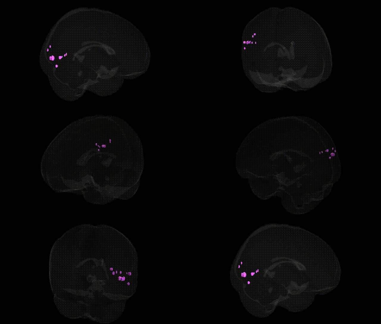
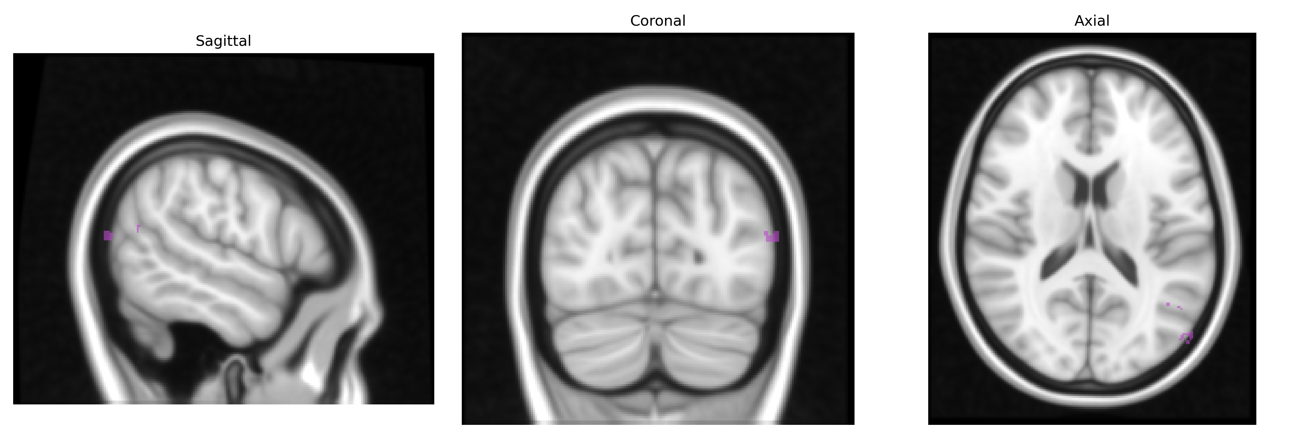
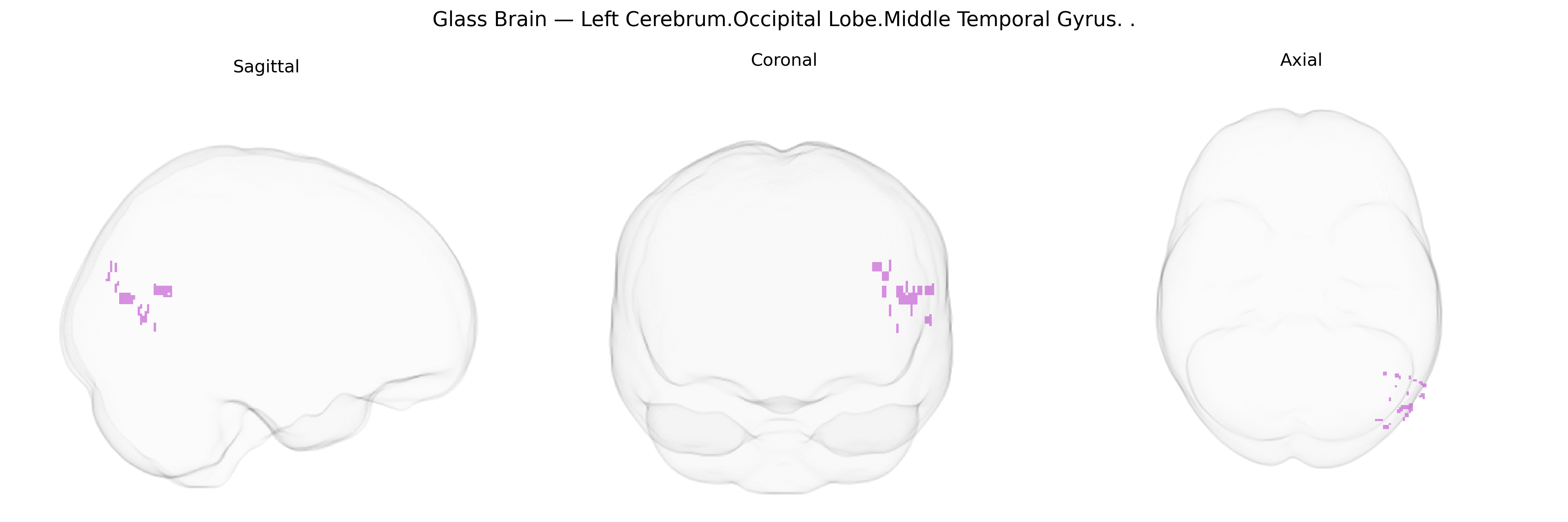
The bilateral Left Cerebrum.Occipital Lobe.Middle Temporal Gyrus, as defined in the Talairach 1 mm atlas, refers to portions of the middle temporal gyrus that extend into or border the occipital lobe region of the left cerebral hemisphere, lying along the lateral temporal–occipital junction. This area is part of the temporal neocortex and is implicated in higher-order visual processing, including motion perception and the integration of visual information with semantic and language-related processes, as well as in aspects of audiovisual integration. It receives inputs from occipital visual areas and projects to distributed temporal and parietal association networks involved in object recognition and conceptual knowledge. There is no direct Wikipedia article for this exact atlas label; a related structure is the Middle temporal gyrus.
The bilateral left occipital lobe–middle temporal gyrus region (as defined in the Talairach 1 mm atlas) participates in visual, semantic, and higher-order associative processing, and genetic associations linked to this territory largely come from imaging-genetics and GWAS of cortical structure and function rather than from this label specifically. Large-scale MRI–GWAS consortia (e.g., ENIGMA, UK Biobank) have reported that common variants in genes involved in neurodevelopment, synaptic function, and myelination (including but not limited to loci near HMGA2, MIR-148A, WNT signaling genes, and several chromatin/remodeling genes) are associated with occipital and temporal cortical thickness, surface area, and sulcal morphology in regions overlapping the middle temporal and adjacent occipital cortices. Polygenic risk for schizophrenia, major depressive disorder, bipolar disorder, autism spectrum disorder, and Alzheimer’s disease has been associated with altered structure or activation in lateral temporal and occipital association cortices, implicating distributed genetic risk rather than single genes. Variants in APOE (particularly ε4), CLU, and other Alzheimer’s disease–related loci show associations with cortical atrophy patterns that often include lateral temporal regions, while dyslexia and other reading-related traits show genetic overlap (e.g., DCDC2, KIAA0319 and other neurodevelopmental genes) with networks encompassing the middle temporal gyrus and occipital word-form and motion areas. Functional imaging GWAS and connectome-wide studies further link polygenic scores for cognitive ability and educational attainment to differences in activation and connectivity of occipito-temporal regions, suggesting that a broad set of neurodevelopmental and synaptic genes contributes to inter-individual variability in structure and function of this bilateral left occipital–middle temporal territory, although no single locus is uniquely or exclusively tied to this specific Talairach-defined region.
Overview generated by GPT-4o (2026).
Region ID: 556
Hemisphere: bilateral
Atlas: Talairach labels 1mm
Full Quality Version: Download MP4
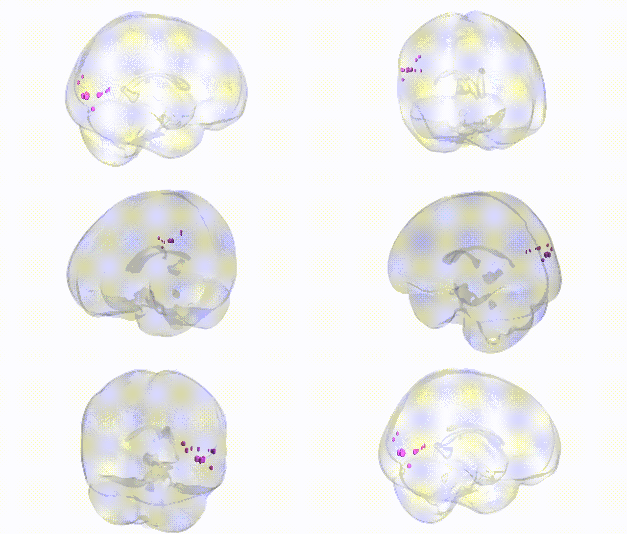
Full Quality Version: Download MP4
Wali Sidiqyar*, Gaurav Rudravaram*, Elyssa M. McMaster, Trent M. Schwartz, Adam M. Saunders, Kurt G. Schilling, Bennett A. Landman "Introducing SPINS: A Shared Public Visualization Library of Neuroanatomical Structures." Medical Imaging with Deep Learning- short paper
This resource is licensed under CC0 1.0 Universal (Public Domain).