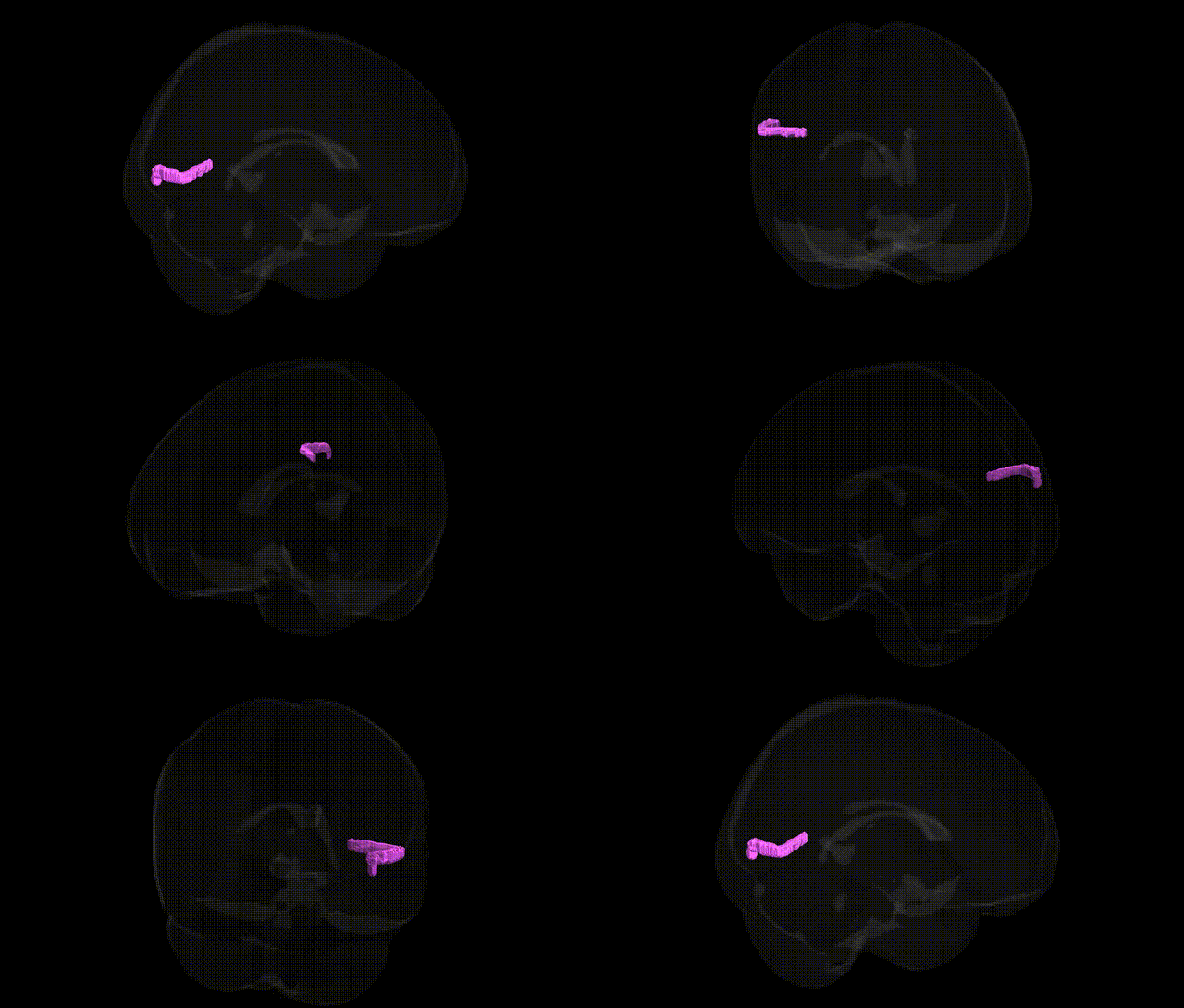
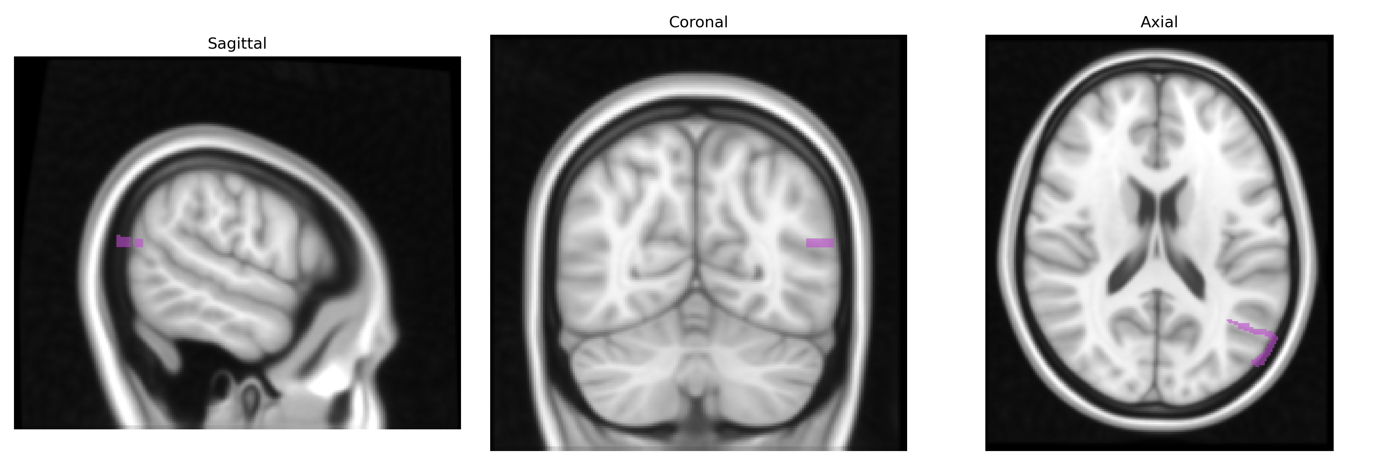
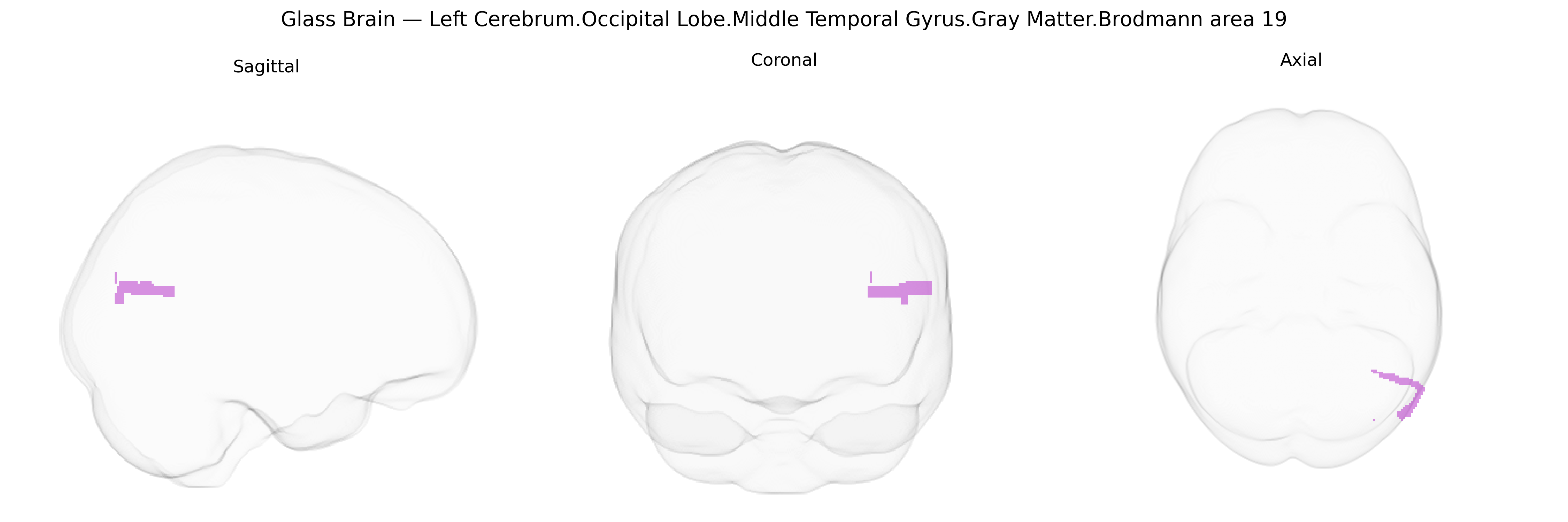
The bilateral Left Cerebrum.Occipital Lobe.Middle Temporal Gyrus.Gray Matter.Brodmann area 19 corresponds to associative visual cortex lying at the intersection of occipital and posterior temporal regions on the left hemisphere, encompassing portions of the middle temporal gyrus that participate in higher-order visual processing. As part of Brodmann area 19, this territory contributes to complex analysis of visual stimuli beyond primary and secondary visual cortices, including integration of form, motion, and object-related features, and interaction with temporal networks involved in visual recognition and semantic processing. Functionally, it belongs to the extrastriate visual cortex and participates in distributed networks supporting visual perception, visuo-semantic integration, and possibly reading-related processes in the dominant hemisphere. There is no direct link for this exact composite region; see the related area Brodmann area 19.
The bilateral left occipital BA19 middle temporal/occipitotemporal region (as labeled in the Talairach 1 mm Atlas) is part of higher-order visual association cortex and has been repeatedly implicated in genetic studies of visual processing, reading and language, and neuropsychiatric risk. Twin and SNP-based heritability analyses of structural MRI consistently show moderate-to-high heritability for gray-matter volume and cortical thickness in BA19 and adjacent occipital-temporal areas, with common variant influences enriched in genes involved in neuronal development, synaptic signaling, and axon guidance (for example, pathways including NRG1–ERBB, semaphorins, and cell-adhesion molecules). Large GWAS of brain imaging traits (ENIGMA, UK Biobank) have identified genome-wide significant loci associated with occipital and occipitotemporal cortical surface area and thickness, including variants near genes such as HMGA2, IGF1, FGFR3, and microtubule- or cytoskeleton-related genes, although mapping to the precise Talairach BA19 middle temporal subregion is approximate given parcellation differences. Functionally, this region overlaps with parts of the visual word form and motion-sensitive areas, and genetic studies of reading and dyslexia (e.g., involving DCDC2, KIAA0319, and ROBO1) as well as language-related GWAS report associations with altered activation or structure in posterior occipitotemporal cortex, including BA19 territories. In psychiatric and neurodevelopmental disorders, polygenic risk for schizophrenia, autism spectrum disorder, and major depression has been associated with subtle thinning or volume differences in occipital and occipitotemporal cortex, and imaging–genetics work links risk loci such as CACNA1C, ZNF804A, and MIR137-related regions to altered connectivity and activation patterns that include BA19. Neurodegenerative and neuroinflammatory conditions, including Alzheimer’s disease, posterior cortical atrophy, and multiple sclerosis, show lesions or atrophy in BA19, and GWAS-identified risk variants (e.g., APOE, CLU, PICALM for Alzheimer’s; HLA and immune-related loci for MS) modulate the degree of gray-matter loss and visual–cognitive impairment affecting this region. Overall, genetic associations for this specific Talairach-defined area are indirect, with most evidence coming from broader occipital or occipitotemporal parcels, but convergent GWAS and imaging–genetics findings indicate that common and rare variants implicated in neurodevelopment, synaptic plasticity, and visual–language processing contribute to individual differences and disease-related vulnerability in this BA19 middle temporal/occipital territory.
Overview generated by GPT-4o (2026).
Region ID: 699
Hemisphere: bilateral
Atlas: Talairach labels 1mm
Full Quality Version: Download MP4
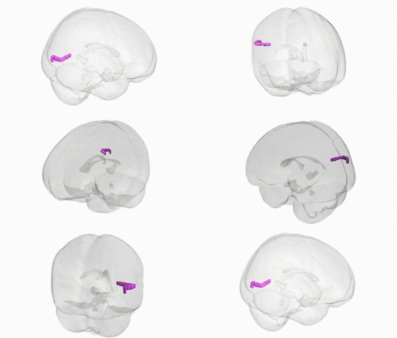
Full Quality Version: Download MP4
Wali Sidiqyar*, Gaurav Rudravaram*, Elyssa M. McMaster, Trent M. Schwartz, Adam M. Saunders, Kurt G. Schilling, Bennett A. Landman "Introducing SPINS: A Shared Public Visualization Library of Neuroanatomical Structures." Medical Imaging with Deep Learning- short paper
This resource is licensed under CC0 1.0 Universal (Public Domain).