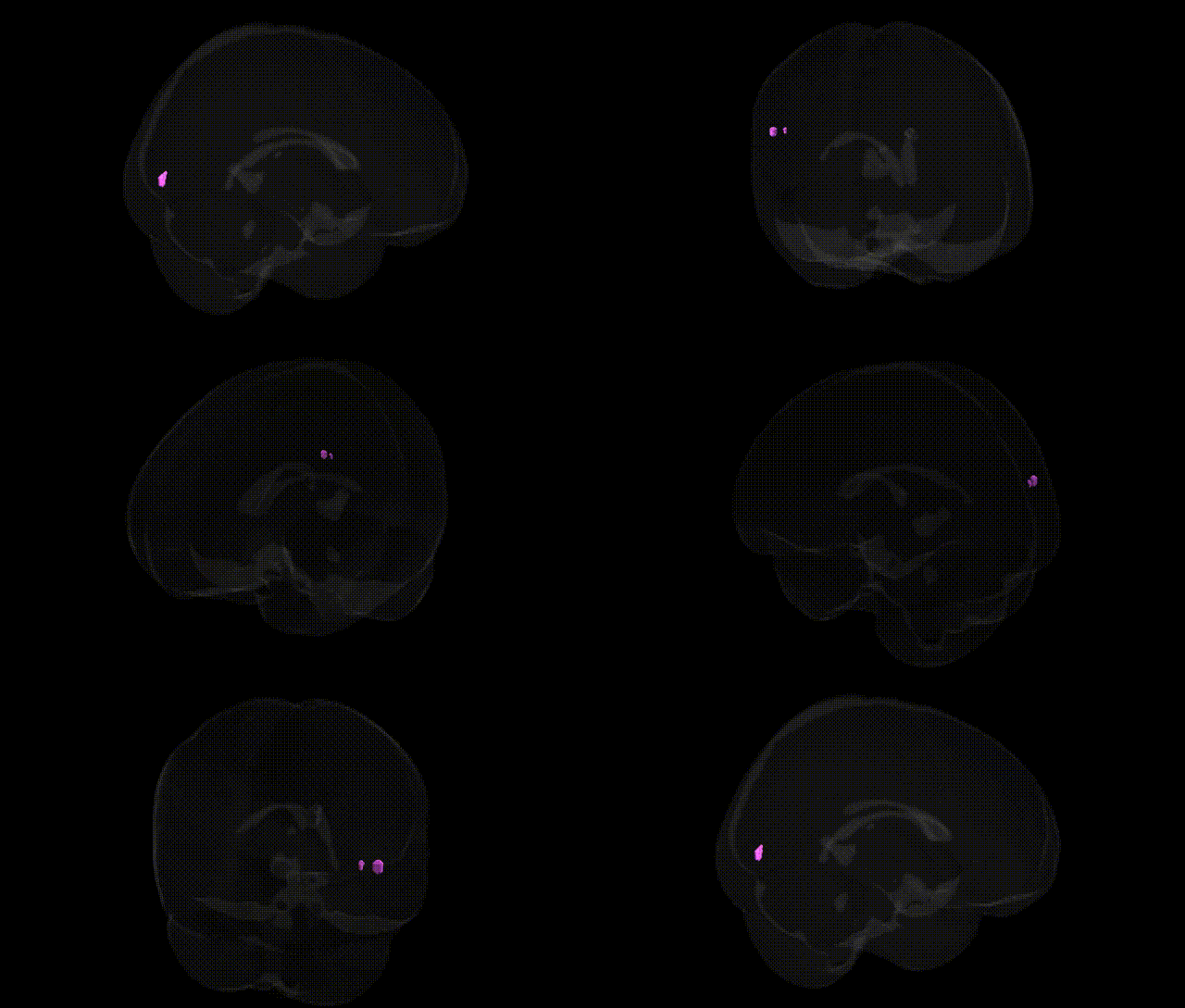
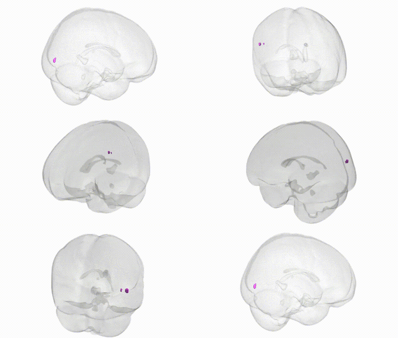
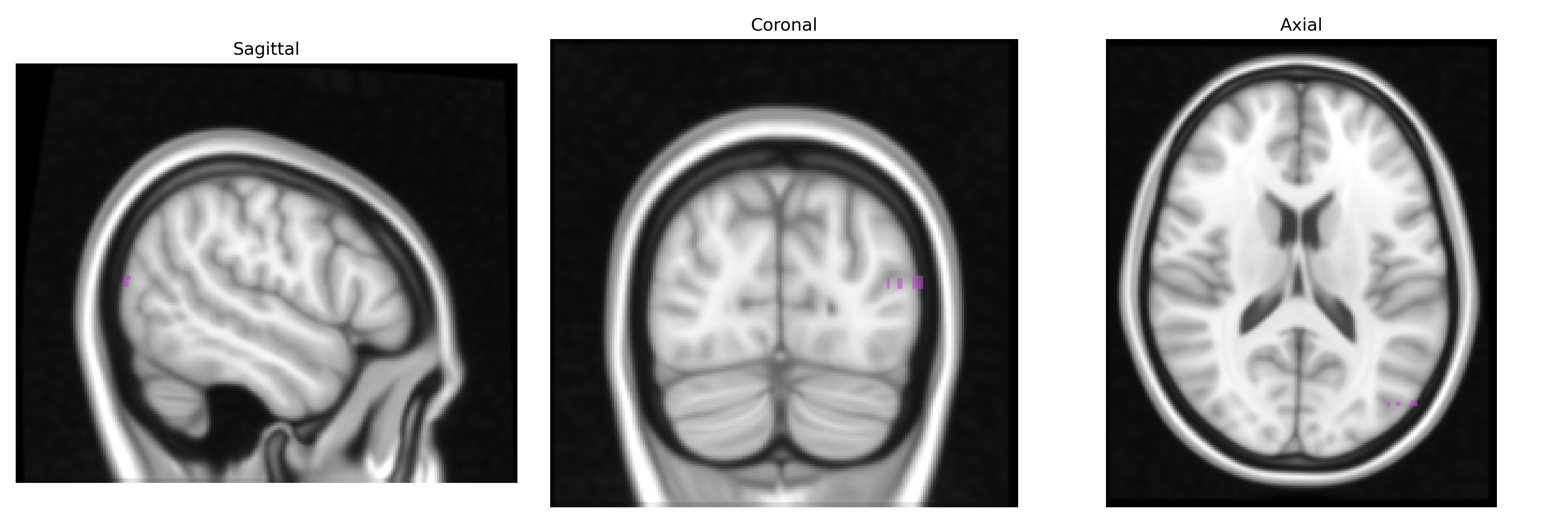
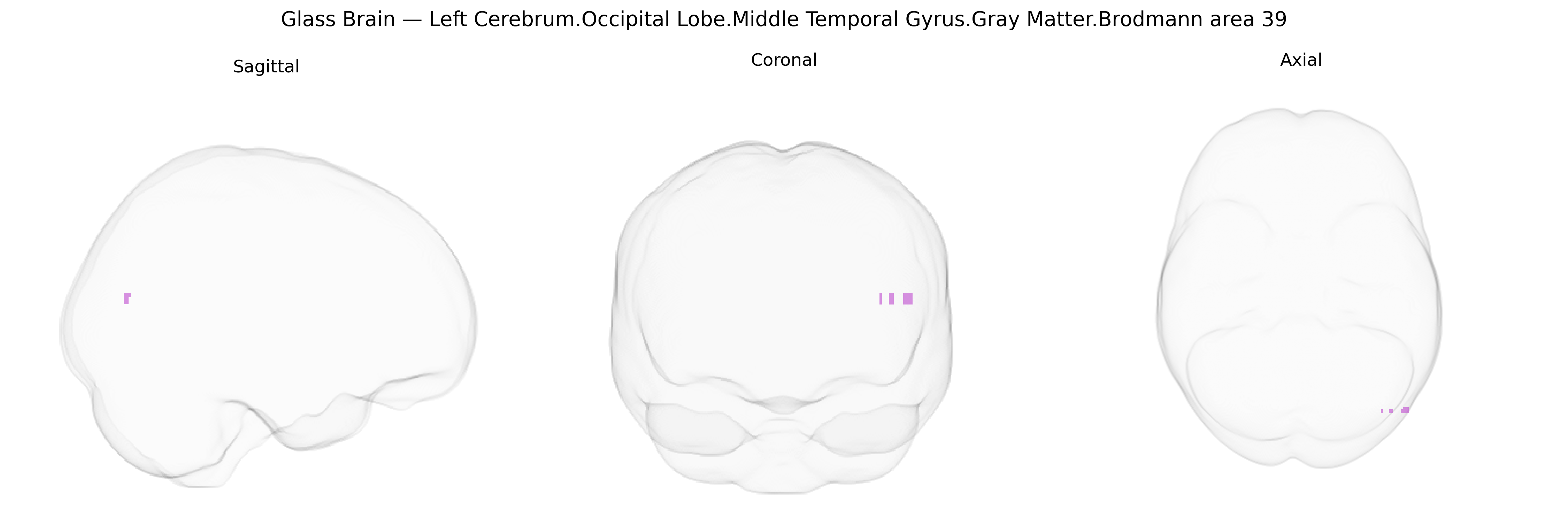
The bilateral Left Cerebrum Occipital Lobe Middle Temporal Gyrus Gray Matter Brodmann area 39 corresponds largely to the angular gyrus region of the inferior parietal/occipitotemporal junction, a multimodal association cortex involved in integrating visual, auditory, and somatosensory information. Cytoarchitectonically defined as Brodmann area 39, this region participates in higher-order language processing (including reading and writing), semantic processing, number cognition, spatial awareness, and aspects of episodic memory retrieval. Functionally, it forms part of the default mode and language networks, receiving convergent input from primary and secondary sensory areas and projecting to frontal association cortices. Lesions in this area are classically associated with Gerstmann syndrome (acalculia, agraphia, finger agnosia, and left–right disorientation) and components of alexia and aphasia, reflecting its role in symbolic and conceptual integration. Brodmann area 39
The bilateral left occipital–temporo‑parietal region corresponding to Brodmann area 39 (angular gyrus/middle temporal–inferior parietal junction in the Talairach 1 mm atlas) has been implicated in multiple genetic and GWAS-based associations, particularly for language, reading, and higher-order cognitive traits. Structural and functional variation in this region has been linked to common variants in genes such as FOXP2, DCDC2, KIAA0319, ROBO1, and CNTNAP2 in studies of developmental dyslexia, specific language impairment, and related language phenotypes, often showing altered gray matter density or activation within BA39. Large-scale ENIGMA and UK Biobank imaging-genetics GWAS have identified associations between cortical thickness/surface area or volume in the angular gyrus/BA39 and loci involving genes related to neurodevelopment (for example MAPT-region variants, microtubule and synaptic genes), as well as polygenic scores for general cognitive ability, educational attainment, and reading performance. BA39 has also emerged in genetic studies of schizophrenia, bipolar disorder, and major depression, where risk alleles in synaptic and calcium-channel genes (e.g., CACNA1C, GRIN2A, complement pathway genes) correlate with altered activation or structure in temporo-parietal association cortex during working memory, language, and default-mode tasks. Additionally, imaging-genetics work in Alzheimer’s disease and primary progressive aphasia links APOE and other AD-risk variants to atrophy and hypometabolism in left temporo-parietal/BA39 regions, consistent with the role of this area in semantic processing and episodic memory networks.
Overview generated by GPT-4o (2026).
Region ID: 702
Hemisphere: bilateral
Atlas: Talairach labels 1mm
Full Quality Version: Download MP4
Full Quality Version: Download MP4
Wali Sidiqyar*, Gaurav Rudravaram*, Elyssa M. McMaster, Trent M. Schwartz, Adam M. Saunders, Kurt G. Schilling, Bennett A. Landman "Introducing SPINS: A Shared Public Visualization Library of Neuroanatomical Structures." Medical Imaging with Deep Learning- short paper
This resource is licensed under CC0 1.0 Universal (Public Domain).