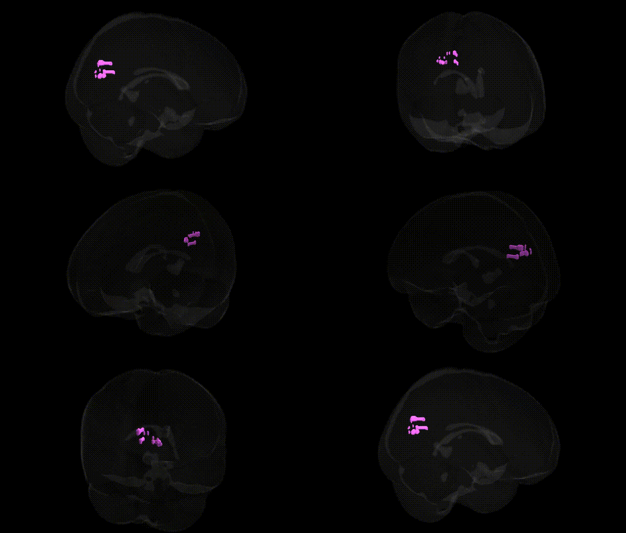
The bilateral Left Cerebrum.Occipital Lobe.Precuneus, as labeled in the Talairach 1 mm atlas, refers to voxels in the left hemisphere’s medial parietal–occipital junction that map to the precuneus region, a key hub within the medial parietal cortex. The precuneus is involved in a broad range of higher-order functions, including visuospatial processing, self-referential and autobiographical memory, aspects of consciousness, and integration of sensory information with internally generated representations. It plays a central role in the default mode network, showing high metabolic activity at rest and deactivation during externally focused tasks. Structurally, it is bordered by the cingulate cortex anteriorly and the occipital cortex posteriorly, and is heavily interconnected with frontal, parietal, and occipital association areas. Precuneus
The bilateral precuneus within the occipital–parietal transition zone (as labeled in the Talairach 1 mm atlas) has been implicated in genetic studies primarily through large-scale neuroimaging GWAS that link common variants to individual differences in cortical thickness, surface area, and connectivity, with notable heritability estimates often exceeding 40–60% for structural measures in this region; genome-wide significant associations have been reported for loci near or within genes such as MIR137, SHANK3, NPTN, and others involved in synaptic development and neuronal signaling, though effects are typically polygenic and small. Imaging–genetics work shows that risk alleles for Alzheimer’s disease (for example, APOE ε4 and CLU variants) are associated with altered precuneus metabolism, connectivity, or atrophy, consistent with the region’s role in early AD pathology, and polygenic risk scores for AD and other neurodegenerative conditions have been tied to structural changes in adjacent medial parietal and occipital cortex. Psychiatric GWAS meta-analyses and imaging–genetics studies indicate that schizophrenia and major depression polygenic risk scores correlate with precuneus volume or default mode network connectivity, implicating genes involved in glutamatergic signaling (e.g., GRIN2A) and synaptic plasticity, while autism spectrum disorder risk variants and polygenic scores have been linked to altered functional coupling of the precuneus with social cognition networks. In addition, genetic variation associated with general cognitive ability, intelligence, and educational attainment (for instance, loci near genes such as MAPT, KIBRA/WFS1, and others from large cognition GWAS) has been related to morphometric and functional differences in the precuneus, consistent with its role in visuospatial integration and self-referential processing. Overall, evidence from GWAS and imaging–genetics supports a highly polygenic architecture in which numerous neurodevelopmental, synaptic, and lipid metabolism genes contribute modestly to the structure and function of the bilateral precuneus, in turn mediating risk for neurodegenerative, psychiatric, and cognitive phenotypes.
Overview generated by GPT-4o (2026).
Region ID: 825
Hemisphere: bilateral
Atlas: Talairach labels 1mm
Full Quality Version: Download MP4
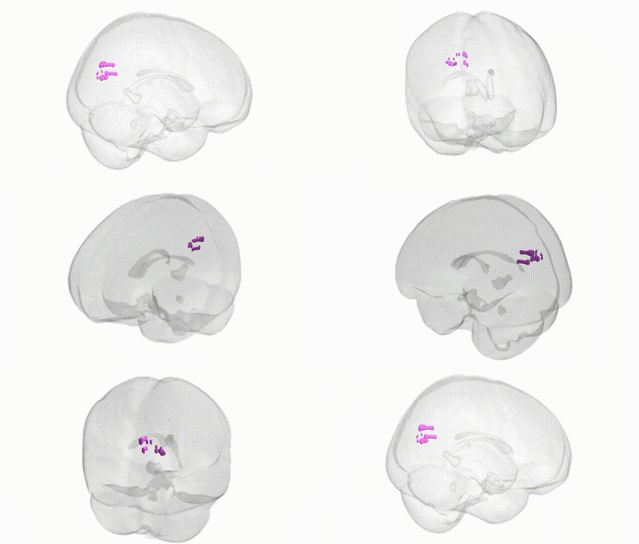
Full Quality Version: Download MP4
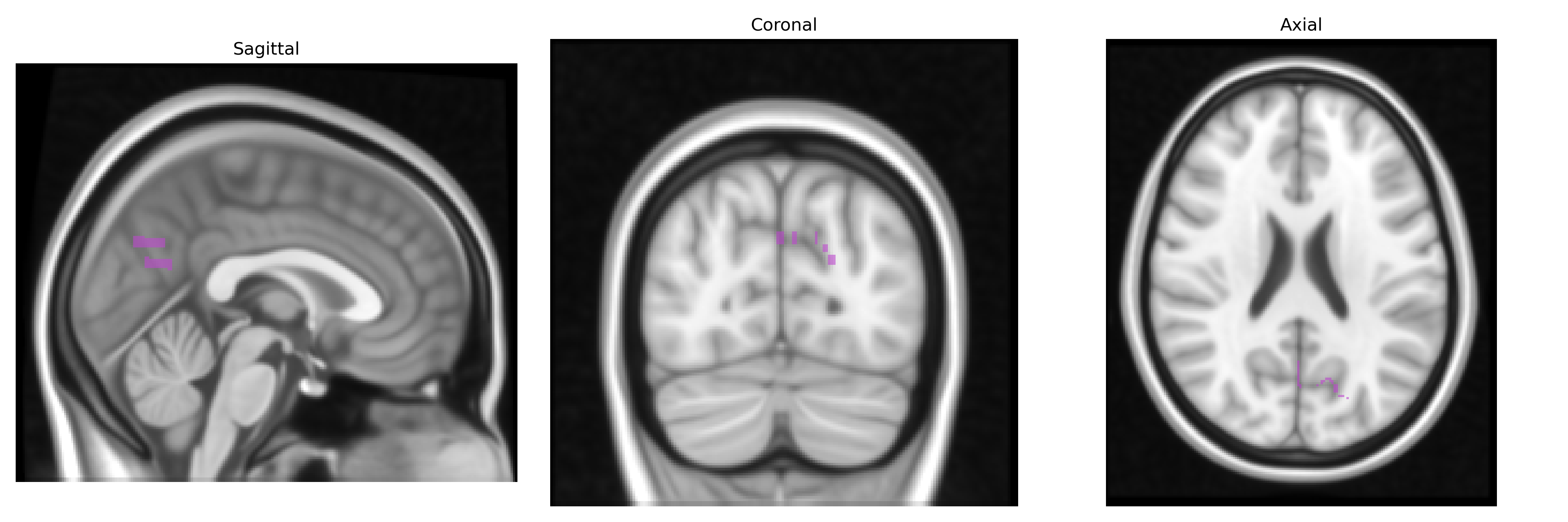
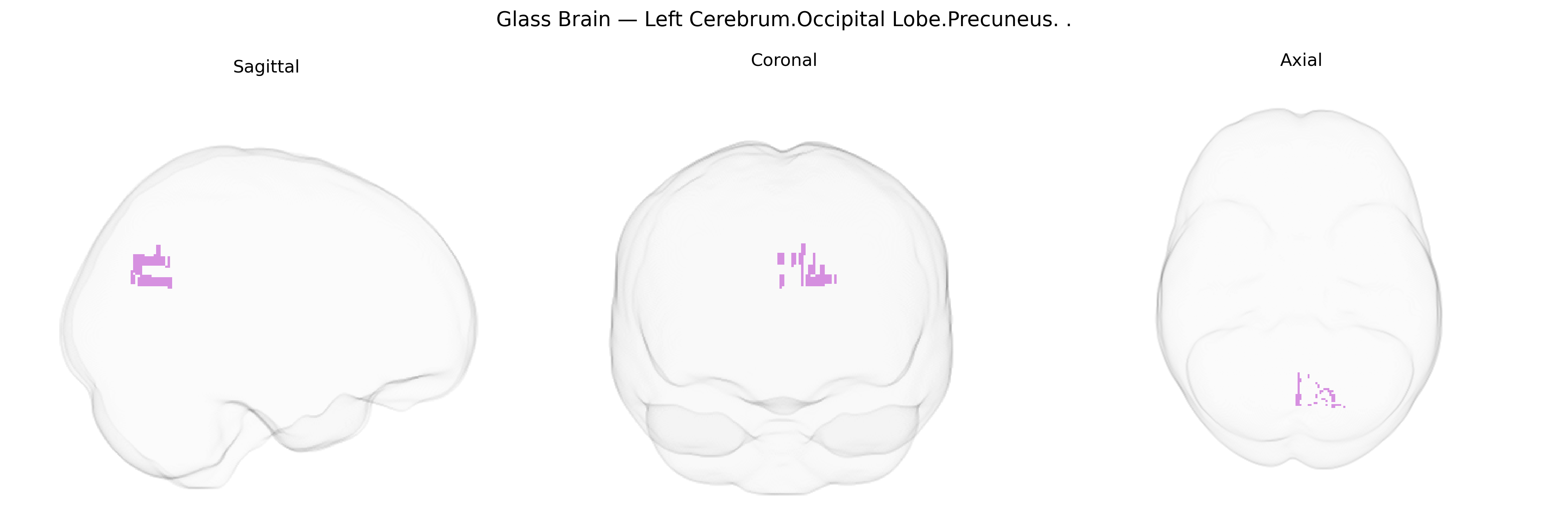
Wali Sidiqyar*, Gaurav Rudravaram*, Elyssa M. McMaster, Trent M. Schwartz, Adam M. Saunders, Kurt G. Schilling, Bennett A. Landman "Introducing SPINS: A Shared Public Visualization Library of Neuroanatomical Structures." Medical Imaging with Deep Learning- short paper
This resource is licensed under CC0 1.0 Universal (Public Domain).