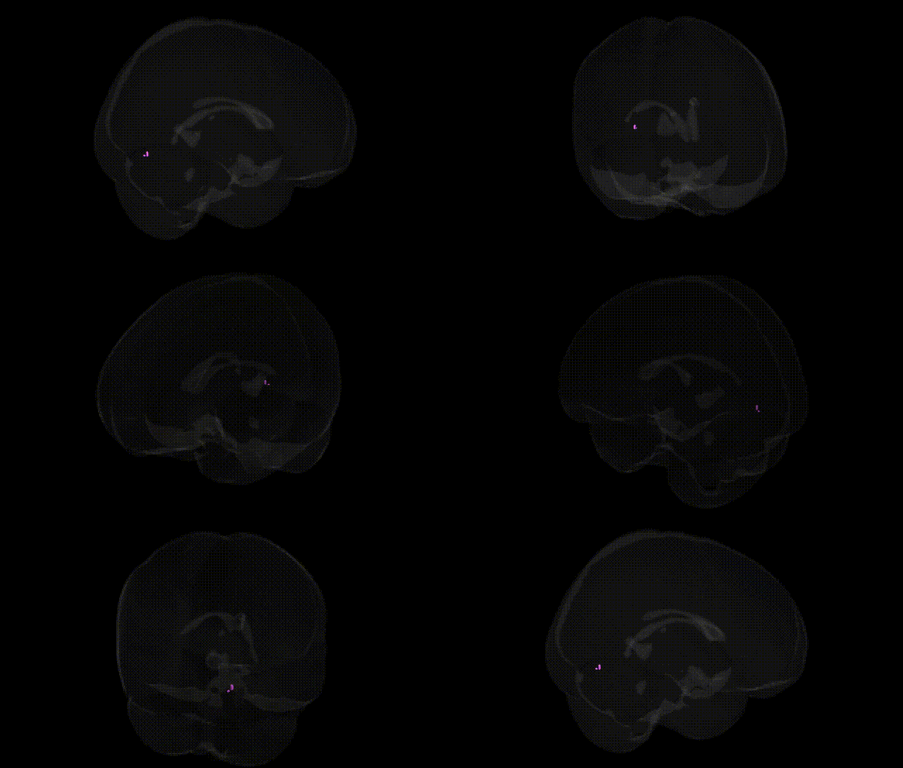
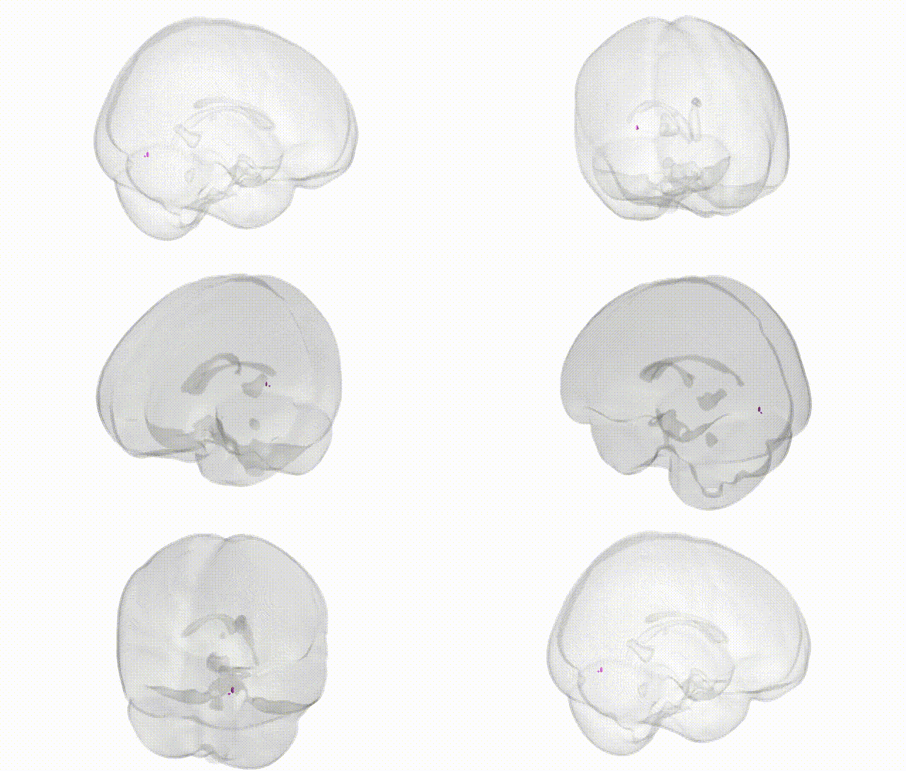
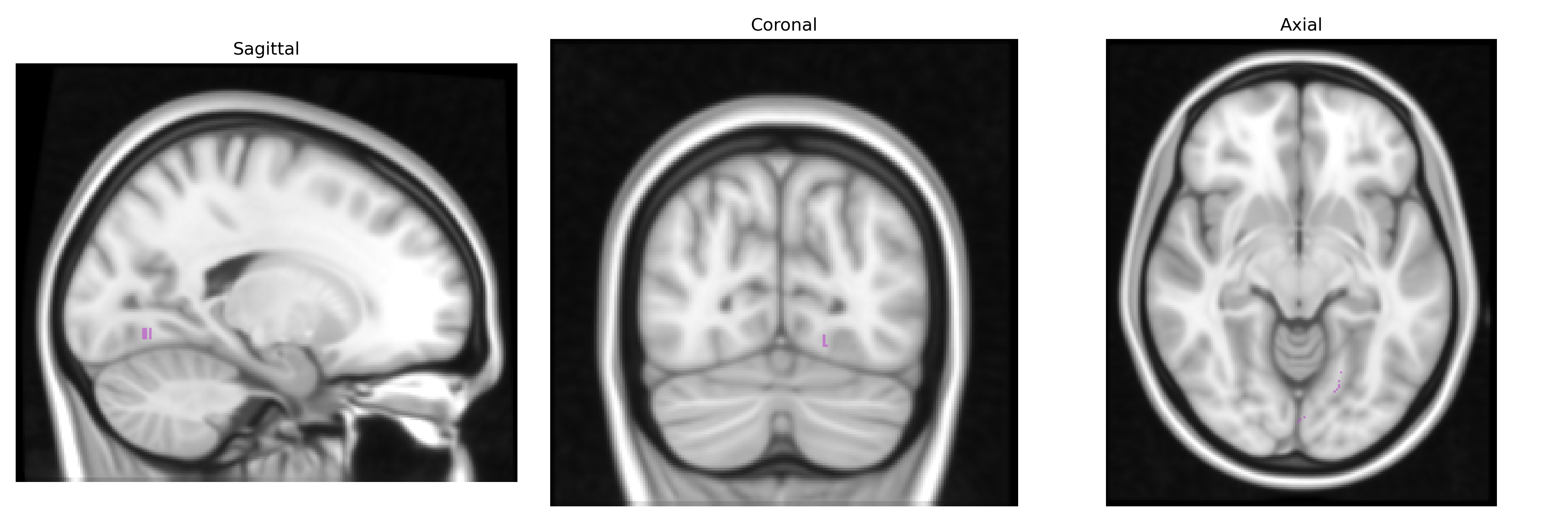
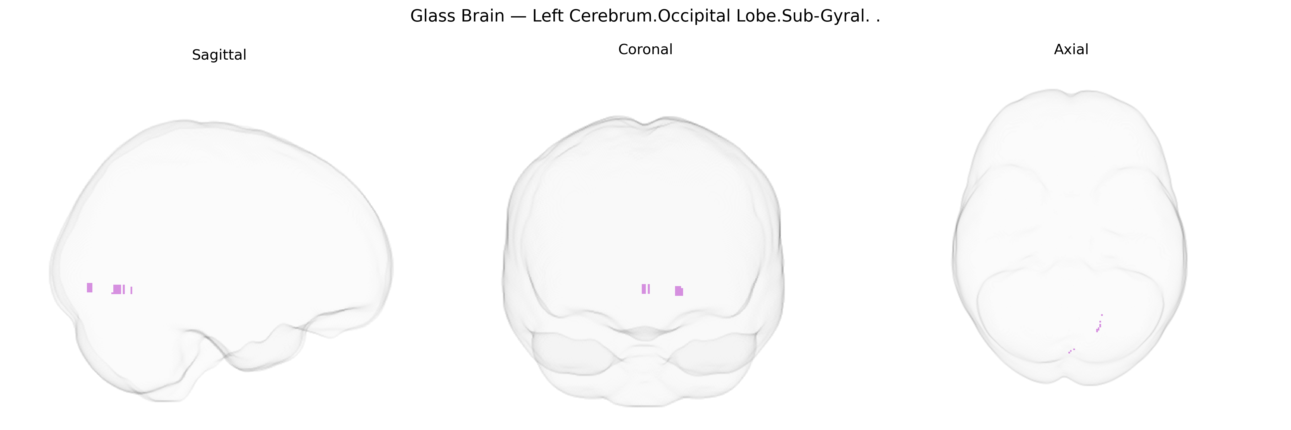
The bilateral Left Cerebrum.Occipital Lobe.Sub-Gyral region, as defined in the Talairach 1 mm atlas, refers to subcortical white matter and immediately subjacent tissue located beneath the cortical gyri of the left occipital lobe, rather than a specific named gyrus or sulcus. This territory contains association and projection fibers that connect primary and higher-order visual cortices with other cortical and subcortical regions, supporting integration, transmission, and coordination of visual information required for form, color, and motion processing. It lies deep to occipital cortical areas such as the cuneus and lingual gyrus and participates in long-range fiber systems (e.g., components of the inferior longitudinal fasciculus) that link the occipital lobe to temporal and parietal regions, thereby contributing indirectly to visual perception, object recognition, and visuospatial functions. There is no direct link for this specific “sub-gyral” label; a related structure description is provided here: Occipital lobe.
The bilateral left cerebrum occipital sub-gyral region (Talairach “Left Cerebrum.Occipital Lobe.Sub-Gyral”) corresponds primarily to deep portions of the primary and associative visual cortices, where genetic associations are typically inferred from imaging–genetics and GWAS of occipital or visual-cortex phenotypes rather than this label specifically. Large-scale brain-structure GWAS (e.g., ENIGMA and UK Biobank) have identified common variants in genes involved in neurodevelopment (such as MAPT, TBR1/TBR2-related transcriptional programs, and WNT/FGF pathway components) associated with occipital cortical thickness, surface area, and volume, with some loci overlapping those implicated in global cortical development and intracranial volume. Polygenic risk for neuropsychiatric disorders, including schizophrenia, bipolar disorder, major depression, and autism spectrum disorder, has been associated with structural and functional variation in occipital subregions, including altered gray-matter volume or cortical thickness in deep occipital areas, suggesting shared genetic architectures between visual-cortex morphology and these conditions. In addition, GWAS of visual acuity, refractive error (myopia), and visual processing traits have reported associations in and around genes affecting axon guidance, synaptic plasticity, and phototransduction that, via imaging–genetic overlap analyses, relate to differences in occipital cortical organization. While no major disorder is defined primarily by pathology restricted to the occipital sub-gyral zone, convergent genetic evidence links this region’s structure and function to broader polygenic influences on cortical development, visual processing, and susceptibility to neurodevelopmental and psychiatric disorders.
Overview generated by GPT-4o (2026).
Region ID: 312
Hemisphere: bilateral
Atlas: Talairach labels 1mm
Full Quality Version: Download MP4
Full Quality Version: Download MP4
Wali Sidiqyar*, Gaurav Rudravaram*, Elyssa M. McMaster, Trent M. Schwartz, Adam M. Saunders, Kurt G. Schilling, Bennett A. Landman "Introducing SPINS: A Shared Public Visualization Library of Neuroanatomical Structures." Medical Imaging with Deep Learning- short paper
This resource is licensed under CC0 1.0 Universal (Public Domain).