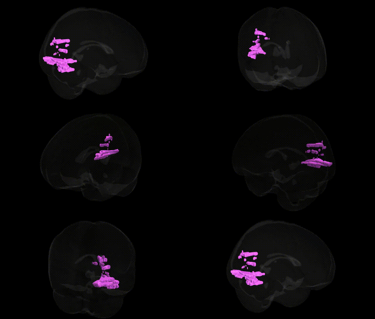
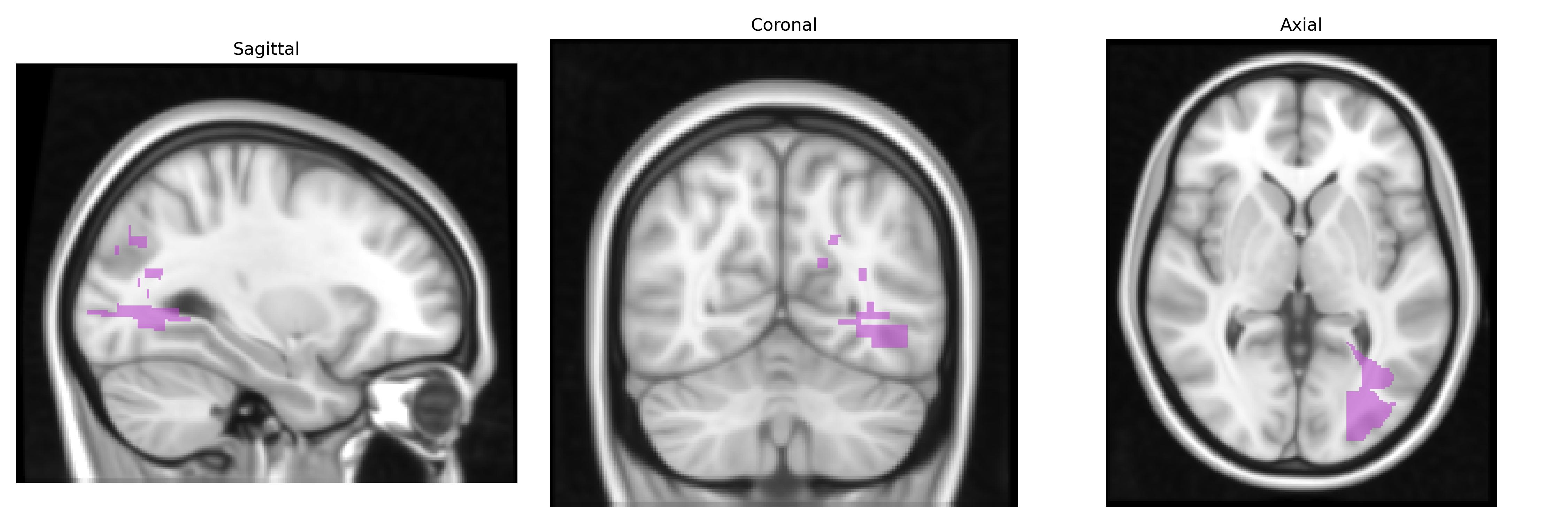
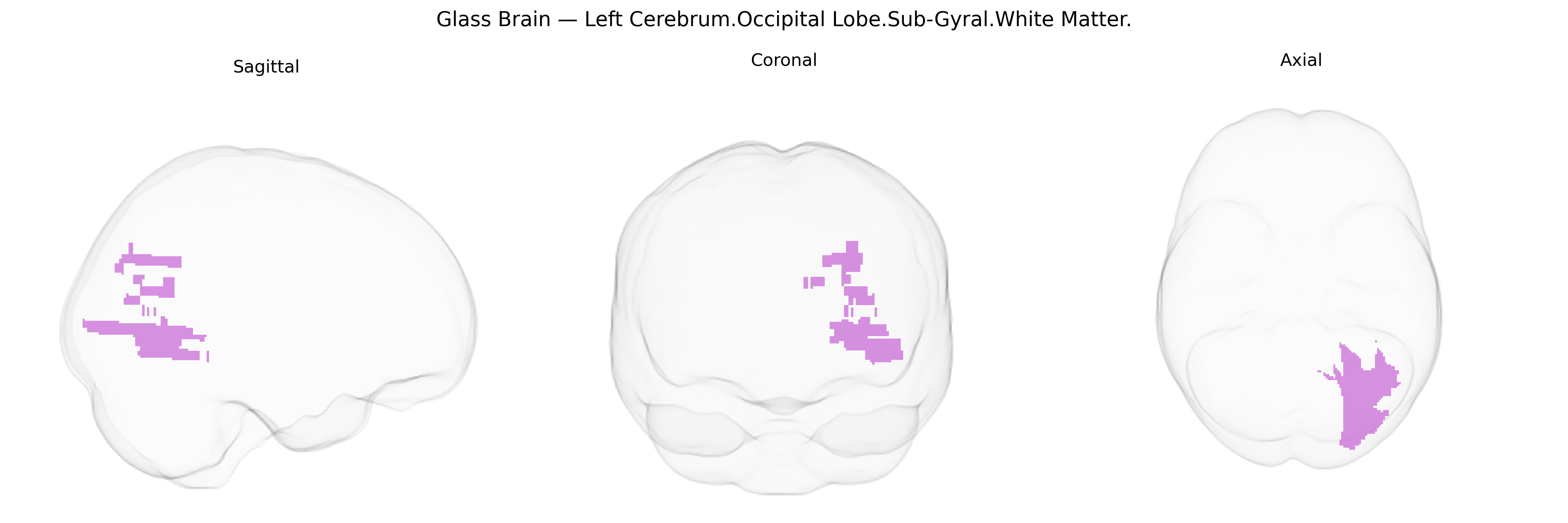
The bilateral Left Cerebrum.Occipital Lobe.Sub-Gyral.White Matter refers to deep white matter tracts located beneath the cortical gyri of the occipital lobe in the left cerebral hemisphere, as defined in the Talairach 1 mm Atlas. This region consists primarily of myelinated axons that interconnect local occipital cortical areas and link them with distant regions, including parietal, temporal, and frontal cortices, as well as subcortical structures. Functionally, these white matter pathways support visual processing by transmitting and integrating information from primary and associative visual cortices, contributing to higher-order visual perception, spatial analysis, and visuomotor coordination. There is no direct Wikipedia article for this specific sub-gyral white matter region; a closely related structure is the Occipital lobe.
The bilateral Left Cerebrum Occipital Lobe Sub-Gyral White Matter, as defined in the Talairach 1 mm atlas, falls within posterior occipital white-matter pathways that contribute to visual and visuospatial processing and has been indirectly implicated in several genetic and GWAS findings through imaging-genetics and voxel-based morphometry studies rather than being a primary, uniquely isolated locus. Large-scale GWAS of brain structure (e.g., ENIGMA and UK Biobank imaging cohorts) have repeatedly identified common variants in genes involved in axonal guidance, myelination, and neurodevelopment—such as NTRK1/2, MAG, MBP, and multiple loci near cell-adhesion and oligodendrocyte-related genes—that influence global and regional white-matter microstructure, including fractional anisotropy and volume within occipital and posterior association tracts. Occipital white matter has been linked to genetic risk for neurodevelopmental and psychiatric disorders, including schizophrenia, autism spectrum disorder, ADHD, and major depression, where polygenic risk scores correlate with altered posterior white-matter integrity; it has also shown susceptibility associations in monogenic or oligogenic leukodystrophies (e.g., variants in PLP1, GFAP, and other myelin genes) that prominently affect occipital and parietal subgyral white matter. In addition, GWAS of visual cortical anatomy and occipital lobe surface area have implicated loci near genes such as PAX6, TBR1, and other neurogenesis regulators, suggesting shared developmental genetic influences on both cortical and immediately subjacent white-matter architecture, while posterior white-matter alterations in genetically mediated conditions such as multiple sclerosis (e.g., HLA-DRB1*15:01 and other immune loci) highlight immune-genetic contributions to vulnerability in this region. Overall, current evidence supports a polygenic architecture in which genes governing neurodevelopment, myelination, and immune function contribute broadly to occipital sub-gyral white-matter structure and its involvement in visual, cognitive, and psychiatric phenotypes, rather than a set of variants uniquely specific to this Talairach-defined region.
Overview generated by GPT-4o (2026).
Region ID: 319
Hemisphere: bilateral
Atlas: Talairach labels 1mm
Full Quality Version: Download MP4
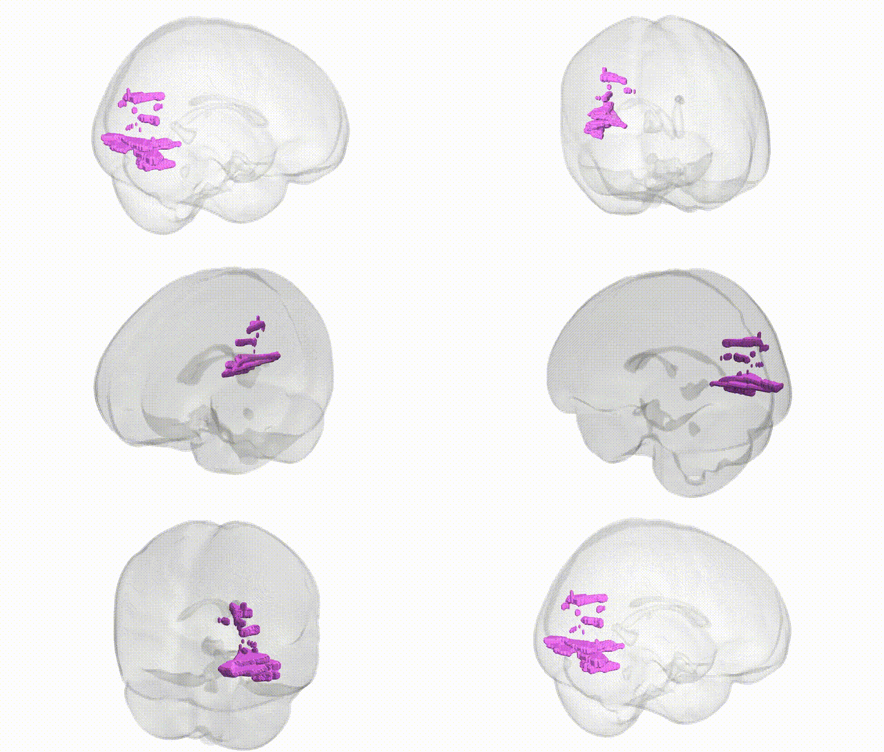
Full Quality Version: Download MP4
Wali Sidiqyar*, Gaurav Rudravaram*, Elyssa M. McMaster, Trent M. Schwartz, Adam M. Saunders, Kurt G. Schilling, Bennett A. Landman "Introducing SPINS: A Shared Public Visualization Library of Neuroanatomical Structures." Medical Imaging with Deep Learning- short paper
This resource is licensed under CC0 1.0 Universal (Public Domain).