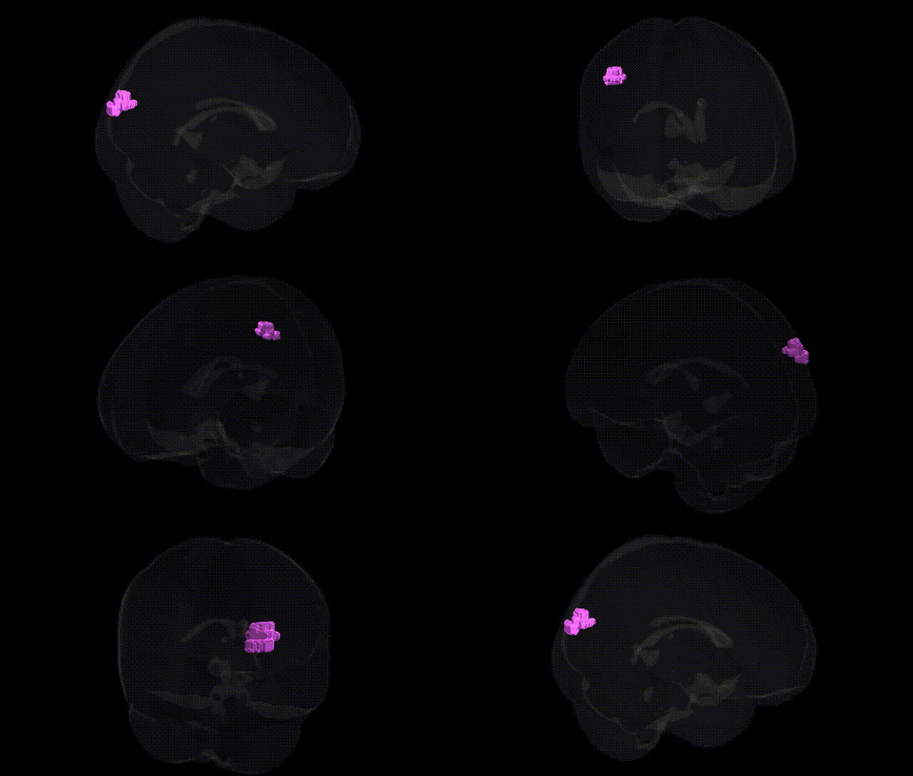
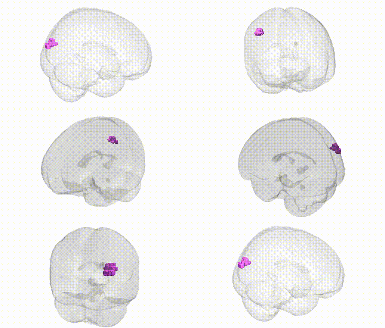
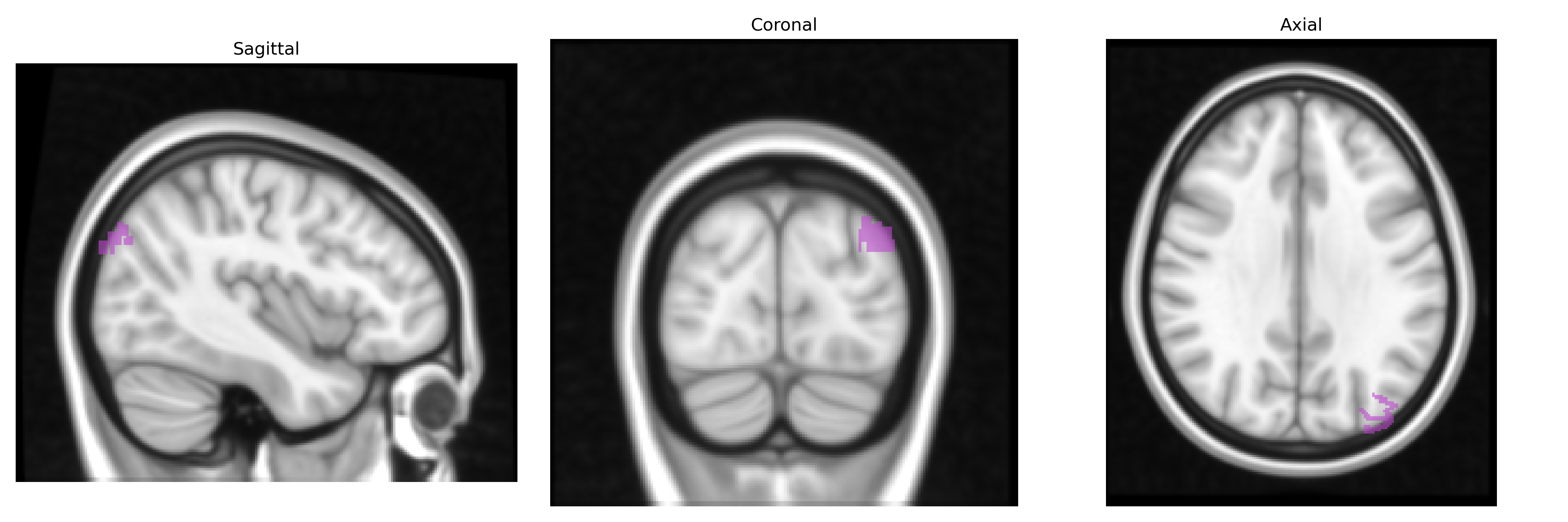
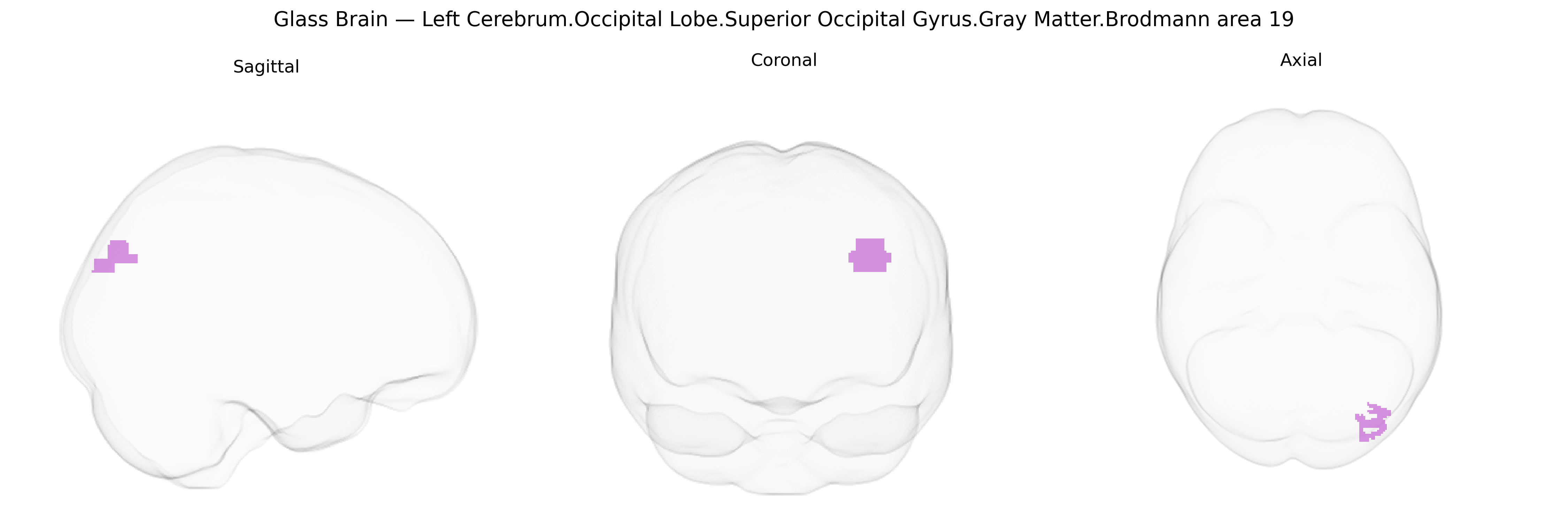
The bilateral Left Cerebrum.Occipital Lobe.Superior Occipital Gyrus.Gray Matter.Brodmann area 19 corresponds to higher-order visual association cortex in the dorsal occipital lobe, located posterior to Brodmann area 18 and extending toward the parietal border, where it participates in the integration of complex visual information. This region contributes to processing of motion, spatial relationships, and object features by integrating inputs from primary and secondary visual cortices, and relaying processed signals to parietal and temporal association areas involved in visuospatial perception, visual attention, and visually guided behavior. Cytoarchitectonically, Brodmann area 19 shows a well-developed granular layer IV and distinct pyramidal layers, reflecting its role in both receiving thalamocortical and corticocortical inputs and sending outputs to widespread association networks. There is no direct link for this exact composite label; a related entry is Brodmann area 19.
The bilateral left occipital superior occipital gyrus gray matter in Brodmann area 19 (BA19), a core component of higher-order visual association cortex, has been implicated in several genetic and GWAS-based findings, although most associations target broader occipital or visual-association regions rather than this exact Talairach-defined parcel. Large structural MRI GWAS (e.g., ENIGMA, UK Biobank) have linked common variants in genes involved in neurodevelopment, synaptic function, and axonal guidance—such as MIR137, CACNA1C, ZNF804A, and genes within the major histocompatibility complex—to variations in occipital cortical thickness and surface area that include BA19, often in the context of schizophrenia, bipolar disorder, and major depressive disorder. Polygenic risk scores for schizophrenia and autism spectrum disorder have shown associations with altered gray-matter volume and connectivity in dorsal visual stream areas encompassing BA19, implicating genes involved in glutamatergic signaling and cortical circuit development. GWAS of imaging-derived phenotypes have identified loci influencing visual cortex metrics (including BA18/19) related to genes regulating neuronal proliferation and myelination (e.g., NRG1/ERBB pathways), while visual-spatial, reading, and face-recognition traits have been tied to genetic variation affecting occipito-temporal and occipito-parietal regions overlapping BA19 (including KIAA0319 and DCDC2 in dyslexia). Additionally, BA19 has appeared in imaging–genetics studies of migraine, multiple sclerosis, and age-related macular degeneration, where risk variants in vascular and inflammatory genes (e.g., in complement pathway genes) correlate with altered occipital activation or atrophy, although these are typically driven by primary eye or systemic pathology with secondary effects on visual association cortex rather than direct BA19-specific genetic determinants.
Overview generated by GPT-4o (2026).
Region ID: 848
Hemisphere: bilateral
Atlas: Talairach labels 1mm
Full Quality Version: Download MP4
Full Quality Version: Download MP4
Wali Sidiqyar*, Gaurav Rudravaram*, Elyssa M. McMaster, Trent M. Schwartz, Adam M. Saunders, Kurt G. Schilling, Bennett A. Landman "Introducing SPINS: A Shared Public Visualization Library of Neuroanatomical Structures." Medical Imaging with Deep Learning- short paper
This resource is licensed under CC0 1.0 Universal (Public Domain).