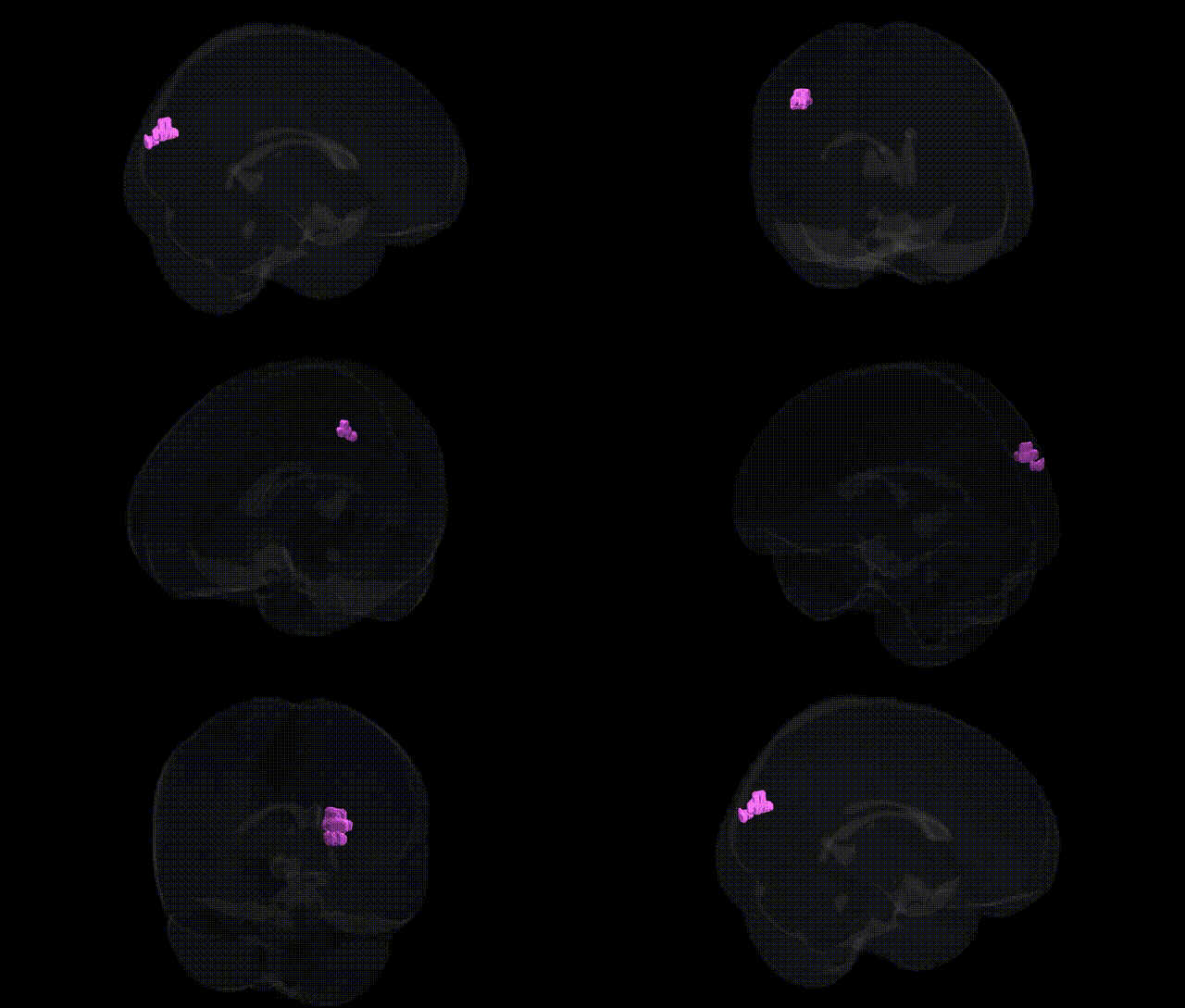
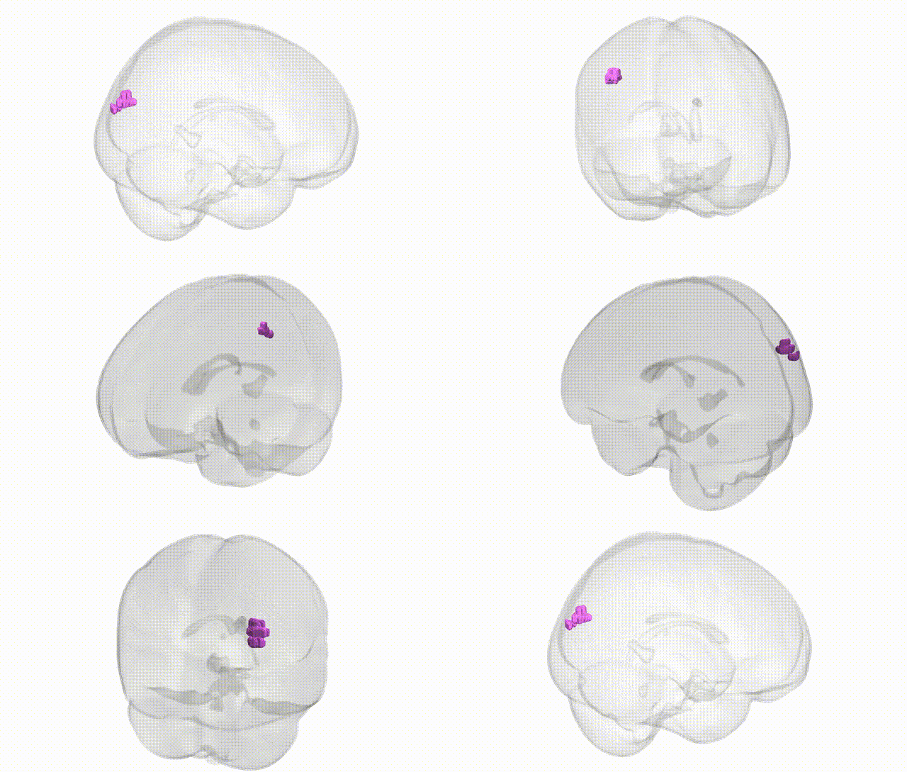
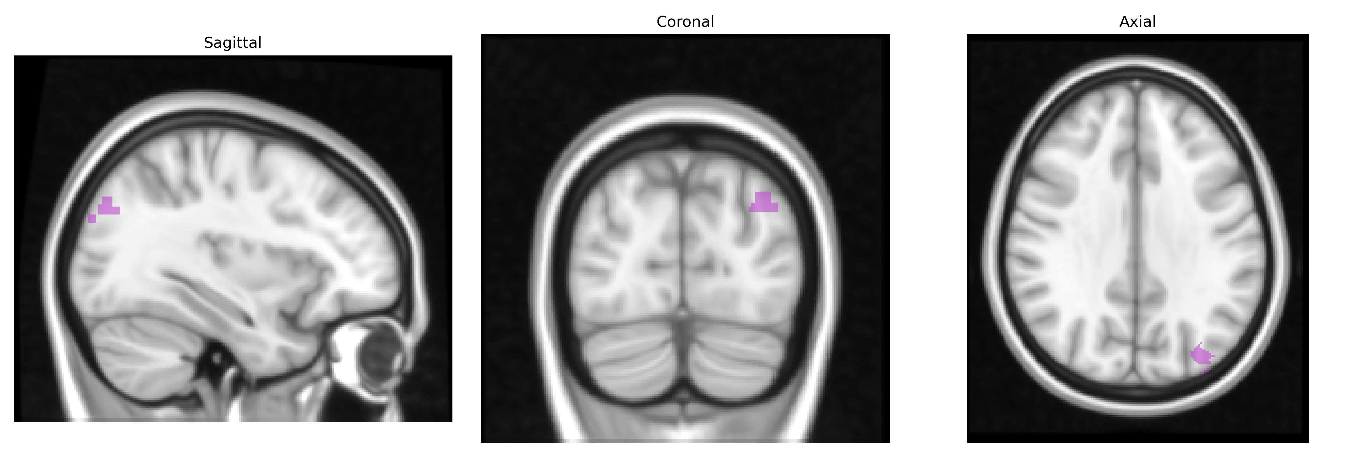
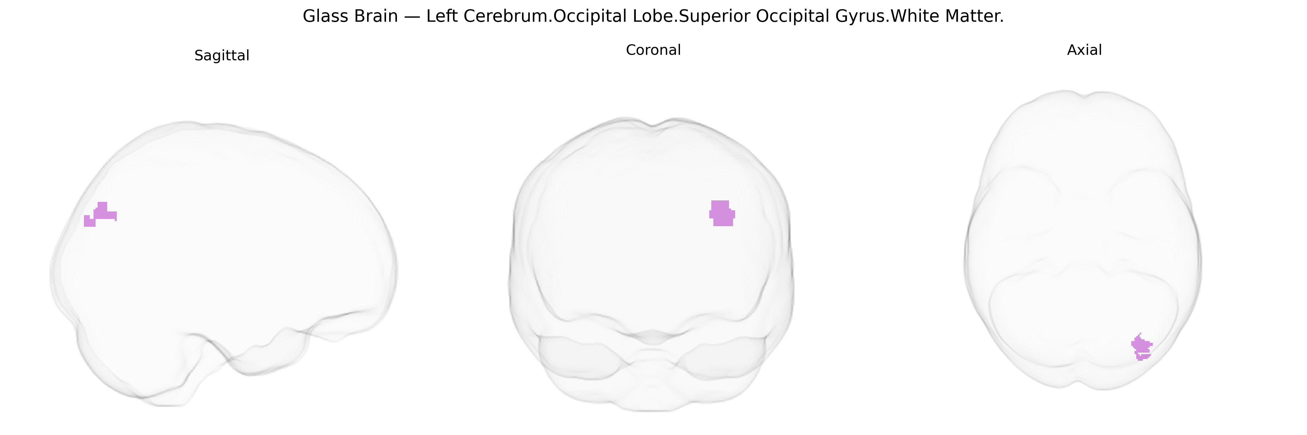
The bilateral Left Cerebrum.Occipital Lobe.Superior Occipital Gyrus.White Matter corresponds to the subcortical myelinated fiber tracts underlying the superior occipital gyrus in the left occipital lobe, as defined in the Talairach 1 mm atlas. These fibers include short association (U-) fibers connecting adjacent occipital gyri, as well as components of longer association pathways such as segments of the superior longitudinal fasciculus and occipito-parietal projections that integrate visual information with dorsal “where/how” processing streams. Functionally, this white matter supports transmission of signals from primary and secondary visual cortices to higher-order parietal and frontal regions involved in visuospatial processing, motion perception, eye–hand coordination, and visually guided attention. Lesions in this area can contribute to higher-order visual deficits, including impairments in spatial orientation and visual attention rather than primary visual loss. There is no direct Wikipedia article for this specific white matter region; see the related structure Occipital lobe.
The bilateral left superior occipital white matter (Talairach 1 mm: Left Cerebrum.Occipital Lobe.Superior Occipital Gyrus.White Matter) has not been the direct focus of many region-specific genetic studies, but diffusion and structural MRI GWAS consistently implicate this territory as part of broader occipital and posterior association white matter tracts. Large imaging-genetics consortia (e.g., ENIGMA, UK Biobank) have reported common variants influencing occipital white matter microstructure—often within or near genes involved in axon guidance (e.g., ROBO1/2, DCC), myelination and oligodendrocyte function (e.g., MAG, MOG, PLP1), and neurodevelopmental patterning (e.g., PAX6, EMX2)—that show effects on fractional anisotropy and mean diffusivity in superior occipital and adjacent parietal pathways. Polygenic risk for schizophrenia, bipolar disorder, and major depression has been associated with reduced integrity of posterior white matter, including superior occipital tracts, while ADHD and autism spectrum disorder risk alleles have been linked to altered occipital connectivity in multimodal imaging-genetics analyses. Occipital white matter metrics overlapping this region also appear in GWAS of general cognitive ability and educational attainment, where variants in genes regulating synaptic plasticity and neuronal connectivity (e.g., CNTNAP2, BDNF-region signals) show small but significant associations with structural connectivity in dorsal visual pathways that traverse the superior occipital gyrus, suggesting that genetic influences on neurodevelopment, myelination, and connectivity contribute to interindividual differences in visual processing, cognition, and vulnerability to neuropsychiatric disorders involving posterior brain networks.
Overview generated by GPT-4o (2026).
Region ID: 850
Hemisphere: bilateral
Atlas: Talairach labels 1mm
Full Quality Version: Download MP4
Full Quality Version: Download MP4
Wali Sidiqyar*, Gaurav Rudravaram*, Elyssa M. McMaster, Trent M. Schwartz, Adam M. Saunders, Kurt G. Schilling, Bennett A. Landman "Introducing SPINS: A Shared Public Visualization Library of Neuroanatomical Structures." Medical Imaging with Deep Learning- short paper
This resource is licensed under CC0 1.0 Universal (Public Domain).