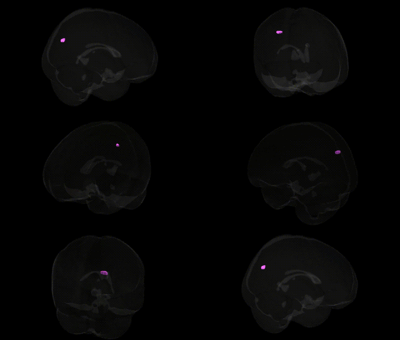
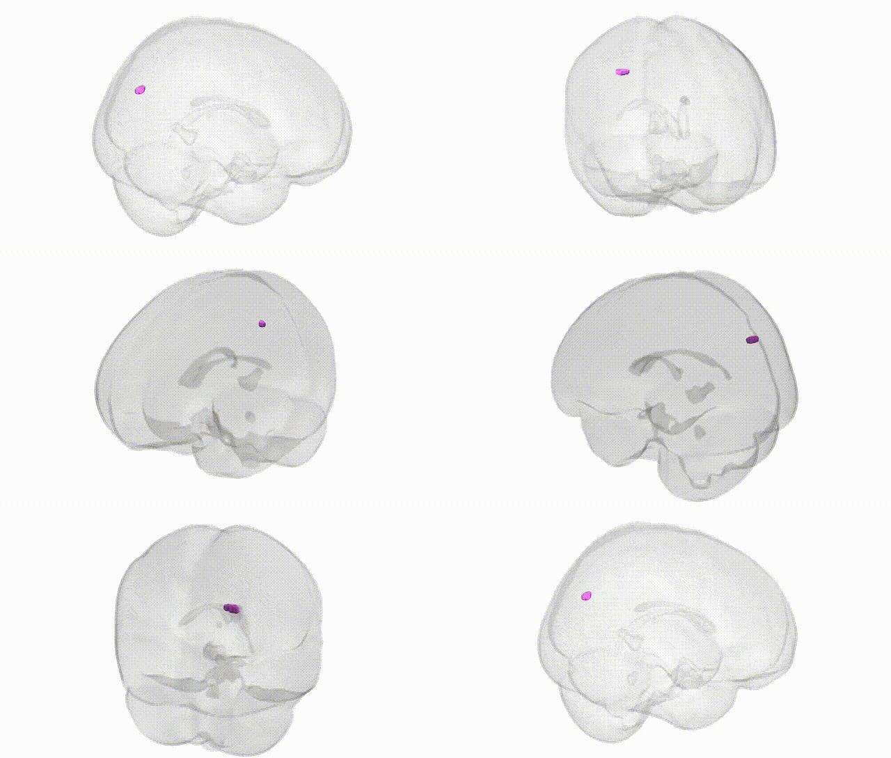
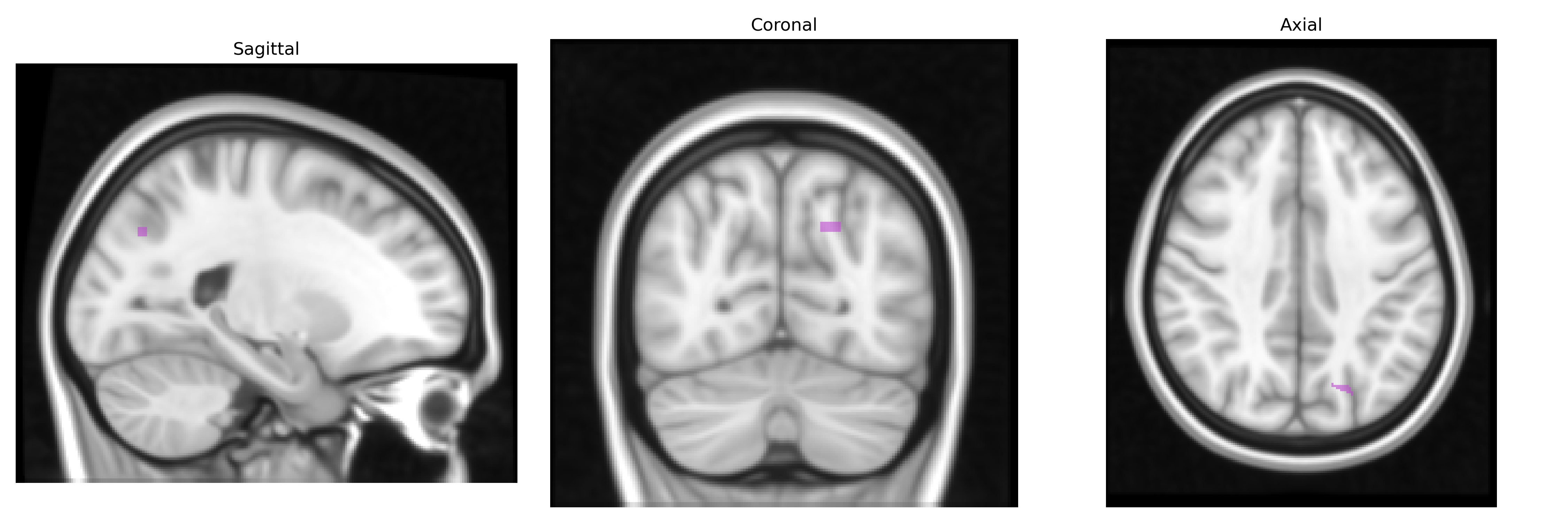
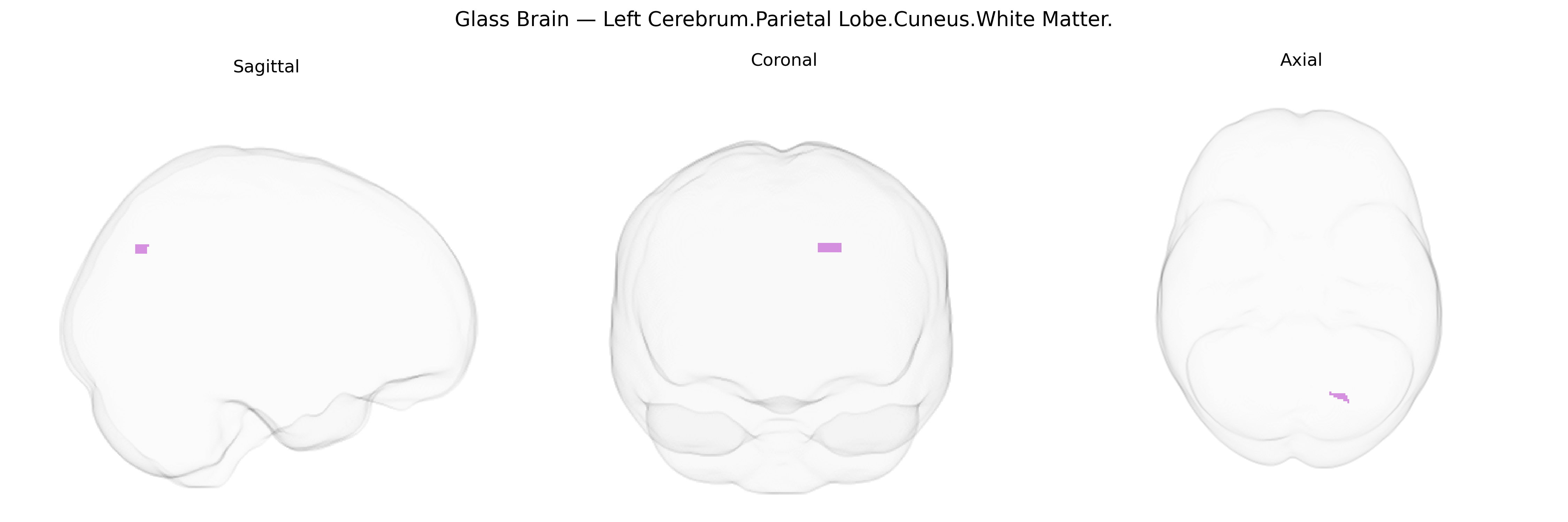
The bilateral Left Cerebrum.Parietal Lobe.Cuneus.White Matter corresponds to the subcortical myelinated fiber pathways underlying the cuneus, a wedge-shaped cortical region on the medial aspect of the occipital lobe near the parieto‑occipital fissure, that participates in primary and associative visual processing. These white matter tracts connect cuneus cortex with adjacent occipital, parietal, and callosal regions, supporting integration of visual information with spatial orientation, attention, and higher-order visuospatial functions. Although the Talairach label places this structure under the parietal lobe, the cuneus is classically described as part of the occipital lobe, emphasizing the atlas-specific nature of this designation. There is no direct Wikipedia article for this exact white matter label; a closely related cortical region is the Cuneus.
The bilateral left parietal cuneus white matter (Talairach 1 mm atlas) has been implicated in genetic studies largely through imaging genetics and GWAS of white‑matter microstructure, cortical connectivity, and neuropsychiatric traits rather than through region‑specific candidate genes. Diffusion MRI–GWAS consortia (e.g., ENIGMA, UK Biobank) have shown that common variants in genes involved in axonal guidance, myelination, and neurodevelopment—such as those in or near NTRK3, CNTNAP2, and ERBB4—contribute to interindividual variation in posterior parietal and occipital white‑matter integrity, which includes the cuneus region and its connections to visual and association cortices. Polygenic risk for schizophrenia, autism spectrum disorder, and major depressive disorder has been associated with altered fractional anisotropy and structural connectivity in parietal–occipital white matter, while Alzheimer’s disease and general cognitive ability (g‑factor) GWAS have linked risk or performance loci (e.g., APOE, CLU, genes in synaptic and lipid‑metabolism pathways) to changes in posterior white‑matter tracts that traverse or border the cuneus. Additional GWAS of visual processing, attention, and reading/spatial abilities suggest that variants affecting posterior association networks influence microstructural properties of this region, though findings are typically reported at the level of large‑scale tracts (e.g., superior longitudinal fasciculus, optic radiations) rather than the cuneus white matter label itself. Overall, genetic associations point to a convergence on neurodevelopmental, synaptic, and myelination pathways shaping the structure and connectivity of the left parietal cuneus white matter and its involvement in cognitive and psychiatric phenotypes.
Overview generated by GPT-4o (2026).
Region ID: 963
Hemisphere: bilateral
Atlas: Talairach labels 1mm
Full Quality Version: Download MP4
Full Quality Version: Download MP4
Wali Sidiqyar*, Gaurav Rudravaram*, Elyssa M. McMaster, Trent M. Schwartz, Adam M. Saunders, Kurt G. Schilling, Bennett A. Landman "Introducing SPINS: A Shared Public Visualization Library of Neuroanatomical Structures." Medical Imaging with Deep Learning- short paper
This resource is licensed under CC0 1.0 Universal (Public Domain).