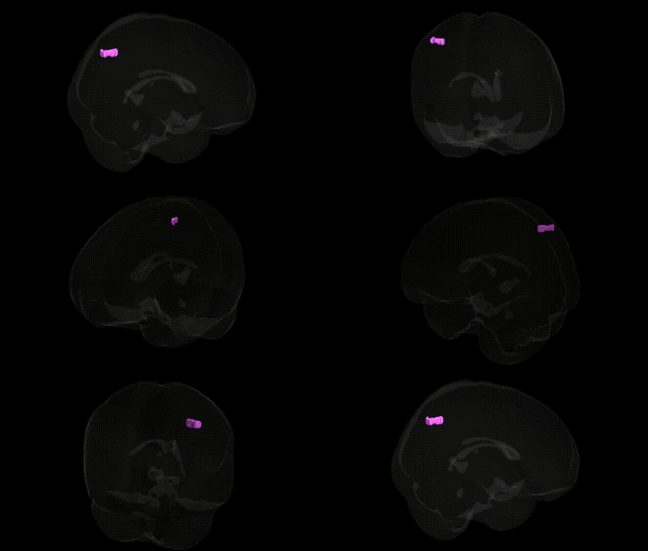
The bilateral Left Cerebrum.Parietal Lobe.Inferior Parietal Lobule.Gray Matter.Brodmann area 7 corresponds to a region of posterior parietal association cortex located just anterior to the occipital lobe and dorsal to the supramarginal and angular gyri, encompassing parts of the superior and inferior parietal lobules in Talairach space. Brodmann area 7 is primarily involved in multimodal integration of visual, somatosensory, and proprioceptive information, contributing to visuospatial processing, spatial attention, hand–eye coordination, and the transformation of sensory input into motor commands for goal-directed actions such as reaching and grasping. Neuronal populations in this area participate in constructing internal representations of extrapersonal space, body schema, and movement planning, with strong connectivity to premotor, motor, and visual association cortices. There is no direct Wikipedia article for this exact composite label; the most closely related structure is Brodmann area 7.
Genetic associations involving the bilateral left cerebrum parietal lobe inferior parietal lobule (IPL) gray matter in Brodmann area 7, as defined in the Talairach 1 mm atlas, largely emerge from imaging-genetics and GWAS of cortical morphology, cognition, and psychiatric risk rather than from region-specific candidate-gene studies. Large-scale cortical GWAS (e.g., ENIGMA, UK Biobank) have identified common variants in genes involved in neurodevelopment and synaptic function (such as HMGA2, MIR2113, KIAA0586, and loci near MCPH1, COL4A2, and genes in Wnt and Notch pathways) associated with parietal cortical thickness, surface area, and gyrification, which encompass BA7/IPL territories. Polygenic scores for general cognitive ability, educational attainment, and mathematical and visuospatial skills show correlations with IPL structure and activation, consistent with the region’s role in attention, working memory, and numerical processing. In neuropsychiatric GWAS, schizophrenia, bipolar disorder, and major depression risk variants—particularly those influencing synaptic plasticity (e.g., in CACNA1C, GRIN2A, and complement pathway genes)—have been linked to altered IPL volume or connectivity, while autism spectrum disorder and ADHD risk loci show convergent effects on dorsal parietal networks involved in social cognition and attentional control. Neurodegenerative and cerebrovascular risk variants, including APOE ε4 and genes modulating amyloid and tau processing, are associated with atrophy patterns affecting inferior parietal and precuneus regions in Alzheimer’s disease, and loci related to small-vessel disease and white-matter integrity (e.g., in COL4A1/COL4A2) contribute to parietal structural changes seen in vascular cognitive impairment. Overall, the genetic architecture of BA7/IPL reflects a polygenic influence shared with broader parietal and frontoparietal networks, with no single variant uniquely specific to this region but multiple loci jointly shaping its development, morphology, and vulnerability to cognitive and psychiatric disorders.
Overview generated by GPT-4o (2026).
Region ID: 1043
Hemisphere: bilateral
Atlas: Talairach labels 1mm
Full Quality Version: Download MP4
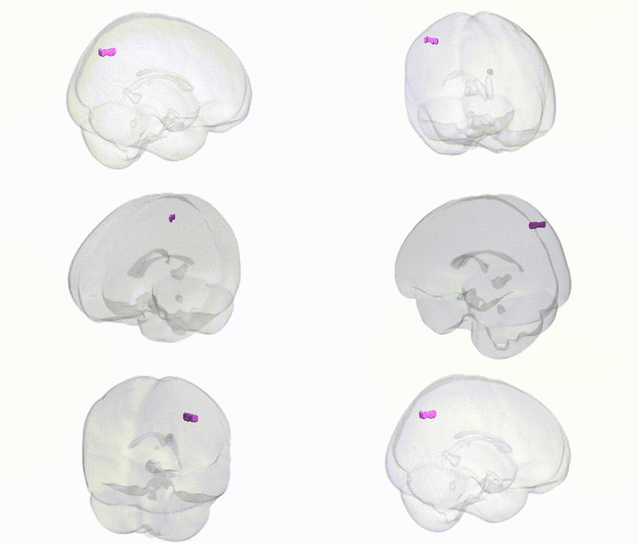
Full Quality Version: Download MP4
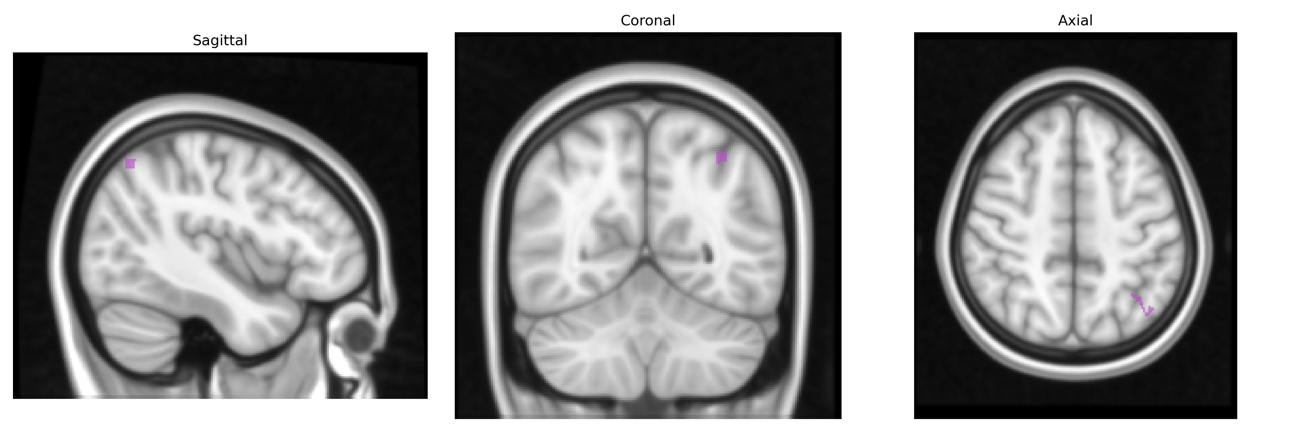
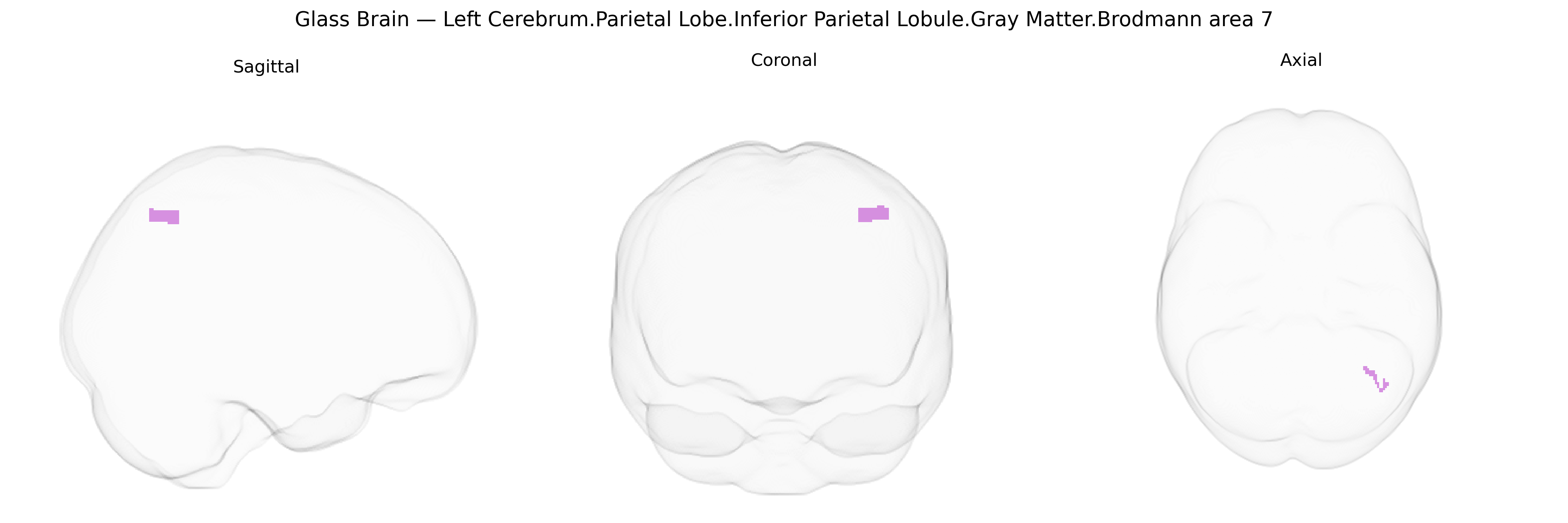
Wali Sidiqyar*, Gaurav Rudravaram*, Elyssa M. McMaster, Trent M. Schwartz, Adam M. Saunders, Kurt G. Schilling, Bennett A. Landman "Introducing SPINS: A Shared Public Visualization Library of Neuroanatomical Structures." Medical Imaging with Deep Learning- short paper
This resource is licensed under CC0 1.0 Universal (Public Domain).