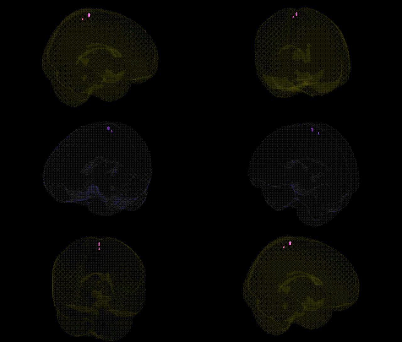
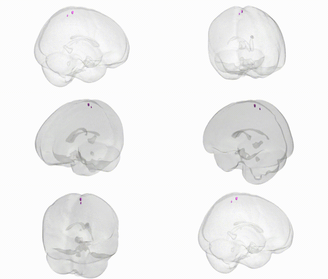
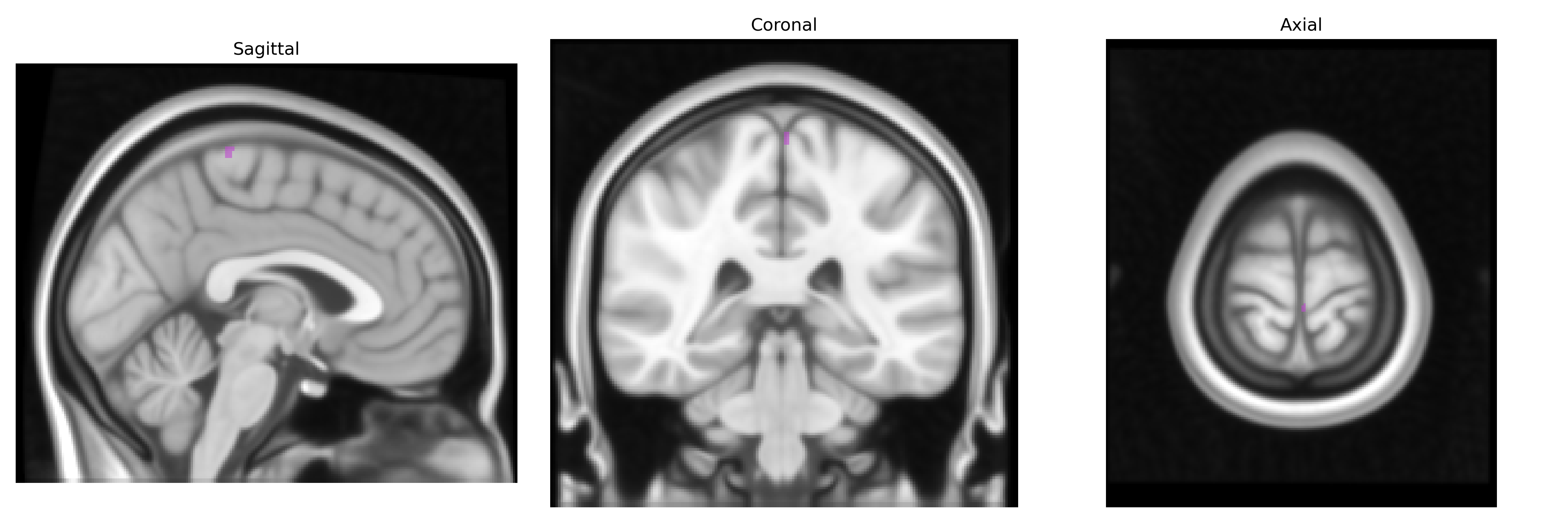
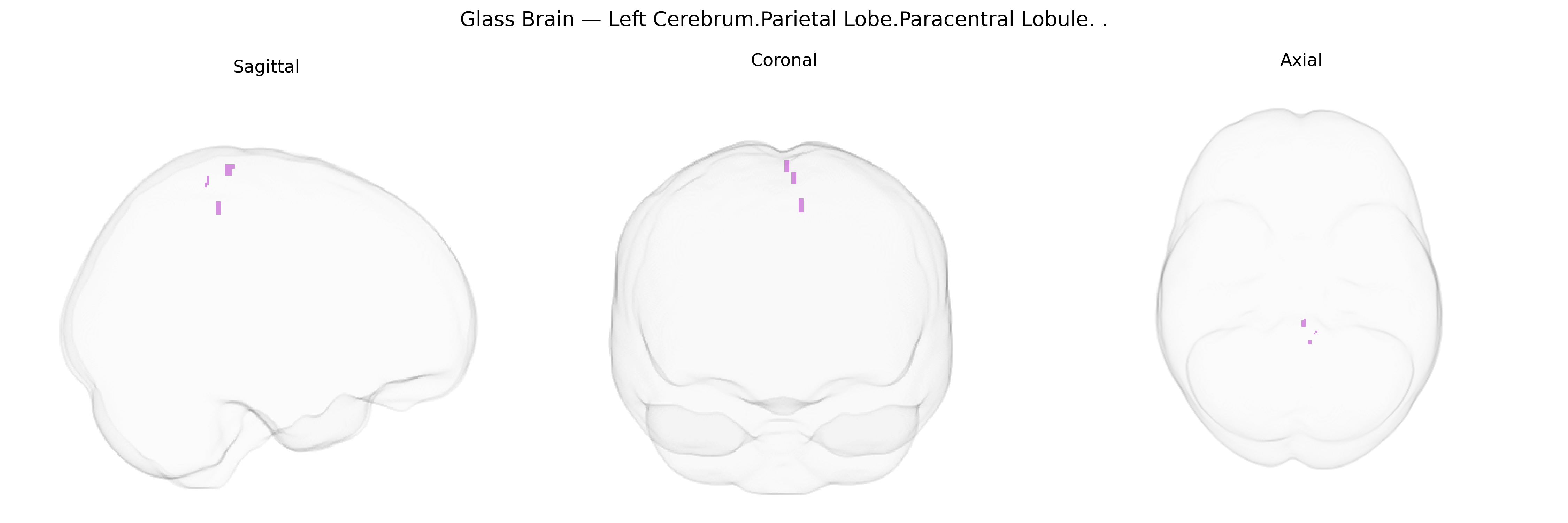
The bilateral Left Cerebrum.Parietal Lobe.Paracentral Lobule corresponds to the paracentral lobule, a medial continuation of the precentral and postcentral gyri that bridges primary motor and primary somatosensory cortices around the medial portion of the central sulcus. It contains somatotopic representations of the contralateral lower limb and perineal regions and participates in voluntary motor control, sensory processing, and aspects of sphincter control for micturition and defecation. Functionally, its anterior part (more frontal) is primarily motor, while the posterior part (more parietal) is primarily somatosensory, and together they integrate motor and sensory information crucial for coordinated movements and bodily awareness of the legs and pelvic region. Paracentral lobule
The bilateral paracentral lobule in the left parietal lobe, as defined in the Talairach 1 mm atlas, has been implicated in multiple genetic and GWAS-based associations, primarily through its roles in motor, somatosensory, and higher-order cognitive networks: structural and functional imaging genetics studies link this region to common variants influencing cortical thickness and surface area (for example, loci near genes such as MAPT, HMGA2, and IGF1 in large ENIGMA and UK Biobank cohorts), with effects on motor and somatosensory cortex morphology; polygenic scores for educational attainment, general cognitive ability, and intracranial volume show associations with paracentral lobule measures, reflecting its integration in sensorimotor–cognitive networks. GWAS of neurological and psychiatric disorders—including multiple sclerosis, amyotrophic lateral sclerosis, Parkinson’s disease, schizophrenia, major depressive disorder, and ADHD—have identified risk loci that, when examined via imaging genetics or lesion mapping, show downstream effects on paracentral lobule structure or activation, especially in relation to motor deficits, fatigue, and altered sensorimotor processing; in addition, genetic variants associated with gait, balance, and motor function (for example in HTT, ATXN2, and other movement-disorder genes) are linked to atrophy or dysfunction in paracentral regions in Huntington’s disease and spinocerebellar ataxias. Pain-related GWAS (chronic back pain, neuropathic pain, and migraine) and somatosensory traits have also been associated with altered activation and morphology of the paracentral lobule in carriers of specific risk alleles, suggesting a genetic contribution to individual differences in somatosensory integration; overall, the region serves as a convergence point where polygenic influences on motor control, somatosensory processing, and cognition manifest as measurable differences in structure and function.
Overview generated by GPT-4o (2026).
Region ID: 1071
Hemisphere: bilateral
Atlas: Talairach labels 1mm
Full Quality Version: Download MP4
Full Quality Version: Download MP4
Wali Sidiqyar*, Gaurav Rudravaram*, Elyssa M. McMaster, Trent M. Schwartz, Adam M. Saunders, Kurt G. Schilling, Bennett A. Landman "Introducing SPINS: A Shared Public Visualization Library of Neuroanatomical Structures." Medical Imaging with Deep Learning- short paper
This resource is licensed under CC0 1.0 Universal (Public Domain).