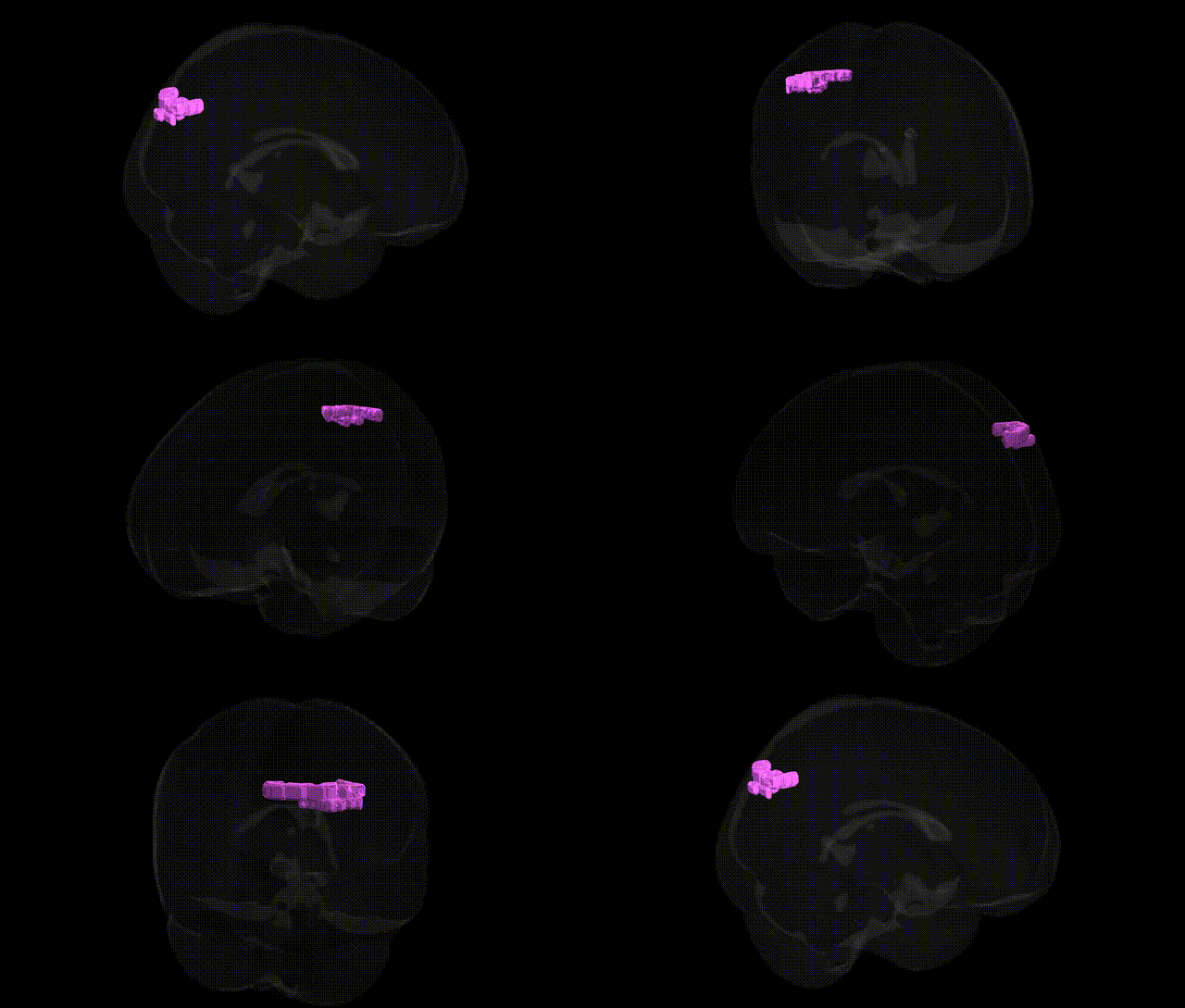
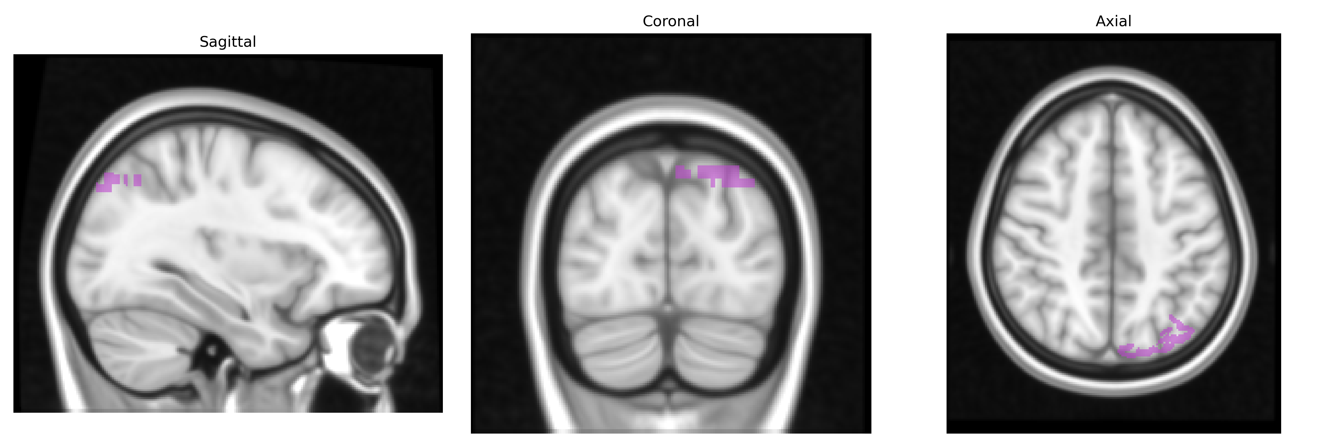
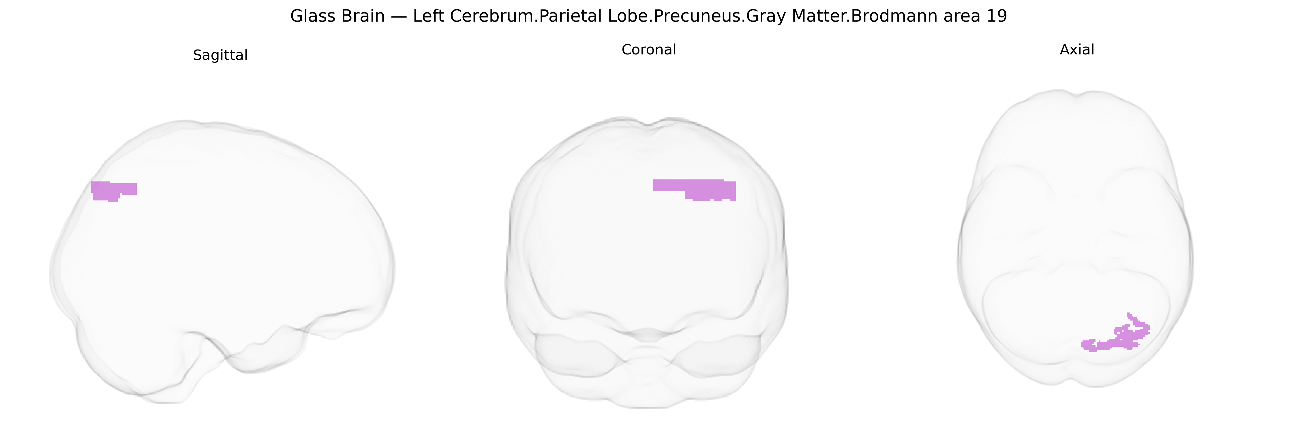
The bilateral Left Cerebrum.Parietal Lobe.Precuneus.Gray Matter.Brodmann area 19 corresponds to association visual cortex located in the medial parietal region, within the precuneus bordering the occipital lobe. As part of Brodmann area 19 (visual association area), this region receives highly processed input from primary and secondary visual cortices and participates in higher-order visual integration, visuospatial processing, mental imagery, and aspects of visuomotor coordination. Functionally, the precuneus portion of BA19 is also implicated in integration of visual information with self-referential and spatial representations, contributing to tasks such as navigation, scene construction, and internally directed cognition. There is no direct link; related area: Brodmann area 19.
The bilateral left precuneus gray matter in Brodmann area 19 (Talairach 1 mm atlas) lies at the intersection of visual association, visuospatial integration, and default-mode network function, and genetic associations involving this region largely emerge from neuroimaging genetics and GWAS of brain structure and connectivity rather than region-specific candidate gene studies. Large-scale GWAS of cortical thickness and surface area have implicated variants in genes related to neurodevelopment and synaptic organization (e.g., pleiotropic loci near HMGA2, MIR9-2, and multiple Wnt and axon-guidance pathway genes) that show effects on parietal and occipital association cortices, including the precuneus and neighboring BA19. Imaging-genetic studies link polygenic risk scores for schizophrenia, major depressive disorder, bipolar disorder, and Alzheimer’s disease to altered precuneus volume, cortical thickness, and functional connectivity, consistent with the role of this region in self-referential processing and episodic memory; several schizophrenia and depression GWAS loci (e.g., near CACNA1C, GRM3, and immune-related genes in the MHC region) show downstream effects on default-mode network hubs that include the precuneus. Additional GWAS of cognitive traits (general intelligence, working memory, and educational attainment) report associations between polygenic load and structural or functional metrics in BA19/precuneus, suggesting that common variants influencing synaptic plasticity and cortical development contribute to individual differences in this region’s morphology and activation. In neurodegenerative disease, APOE ε4 and other Alzheimer’s disease risk loci (CLU, PICALM, BIN1, TREM2) are consistently associated with precuneus hypometabolism and atrophy, reflecting early vulnerability of this hub to amyloid and tau pathology, although these findings are at the regional network level rather than specific to BA19 cytoarchitecture. Overall, genetic associations to this precise bilateral left BA19 precuneus parcel are indirect, derived from broader parietal–occipital and default-mode network analyses, but converge on pathways governing cortical development, synaptic signaling, and neurodegeneration.
Overview generated by GPT-4o (2026).
Region ID: 986
Hemisphere: bilateral
Atlas: Talairach labels 1mm
Full Quality Version: Download MP4
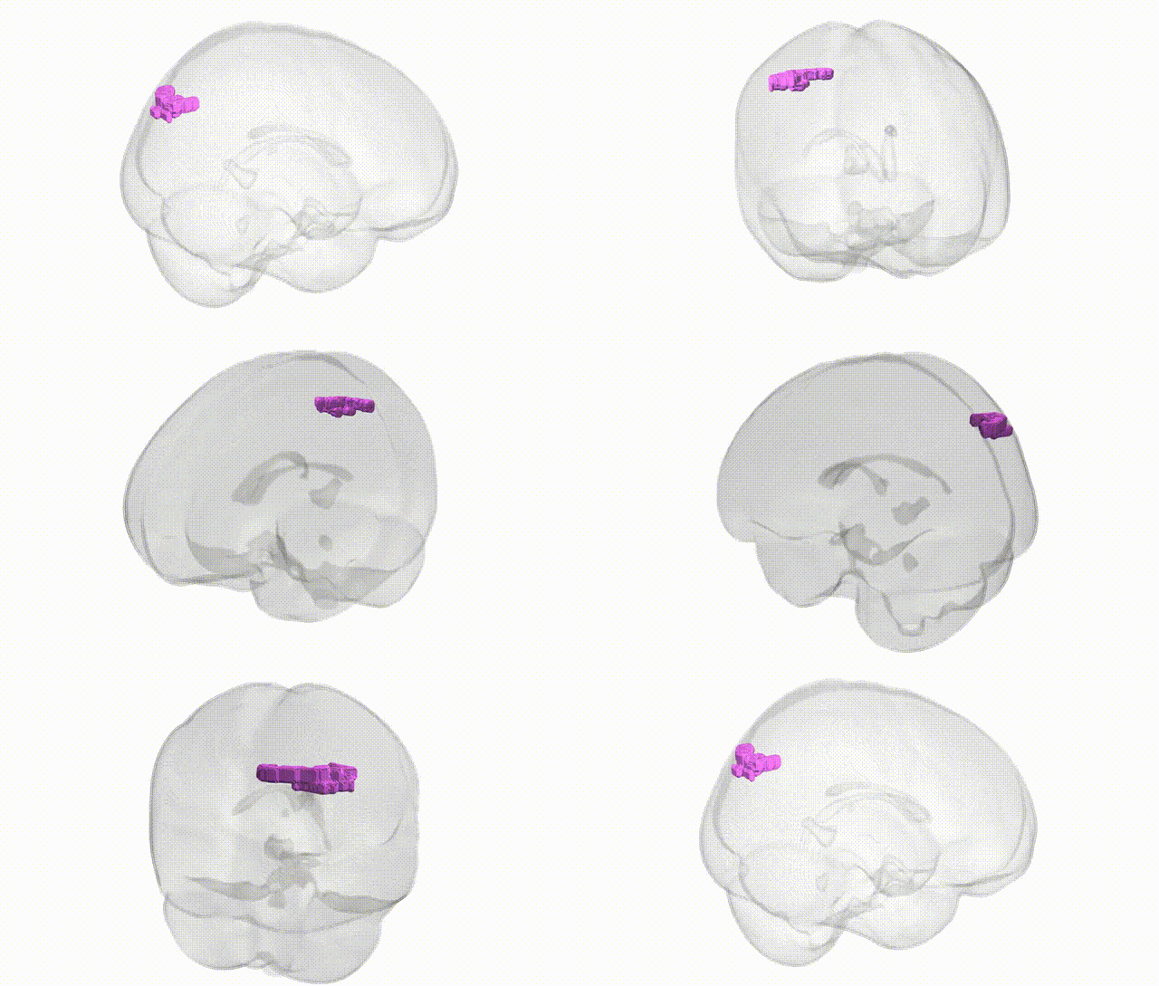
Full Quality Version: Download MP4
Wali Sidiqyar*, Gaurav Rudravaram*, Elyssa M. McMaster, Trent M. Schwartz, Adam M. Saunders, Kurt G. Schilling, Bennett A. Landman "Introducing SPINS: A Shared Public Visualization Library of Neuroanatomical Structures." Medical Imaging with Deep Learning- short paper
This resource is licensed under CC0 1.0 Universal (Public Domain).