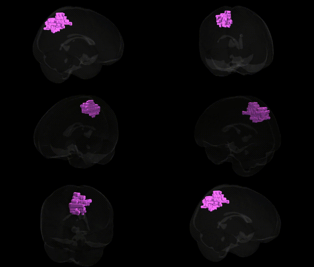
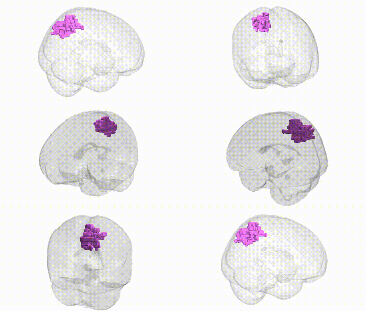
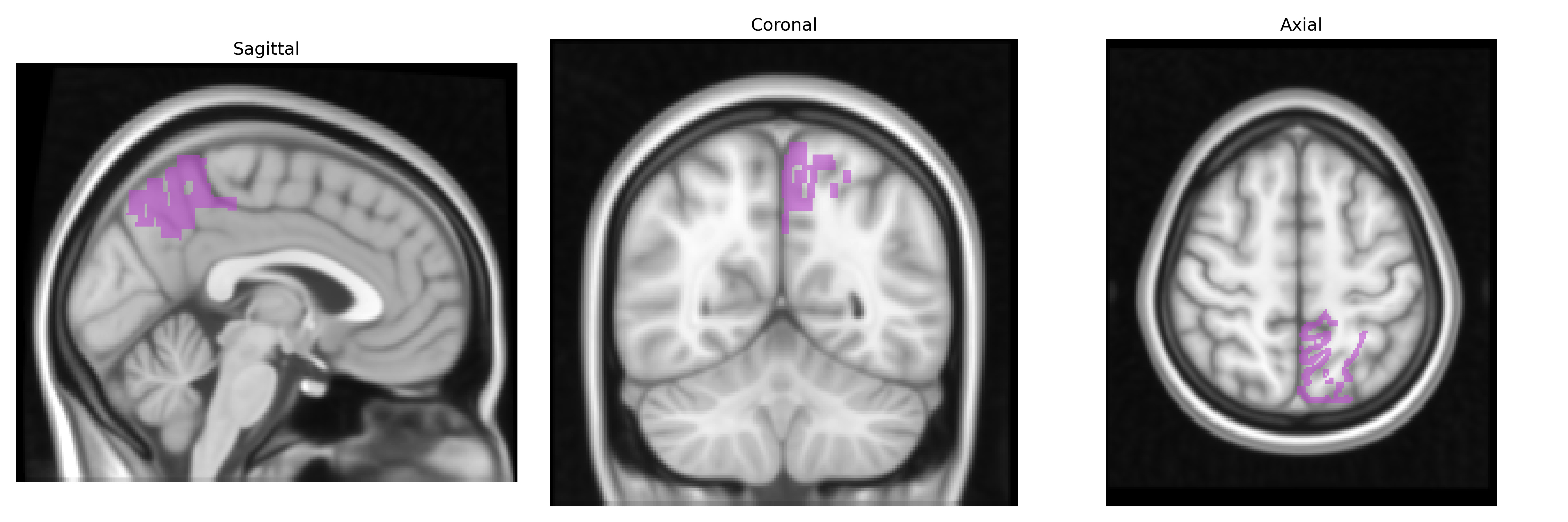
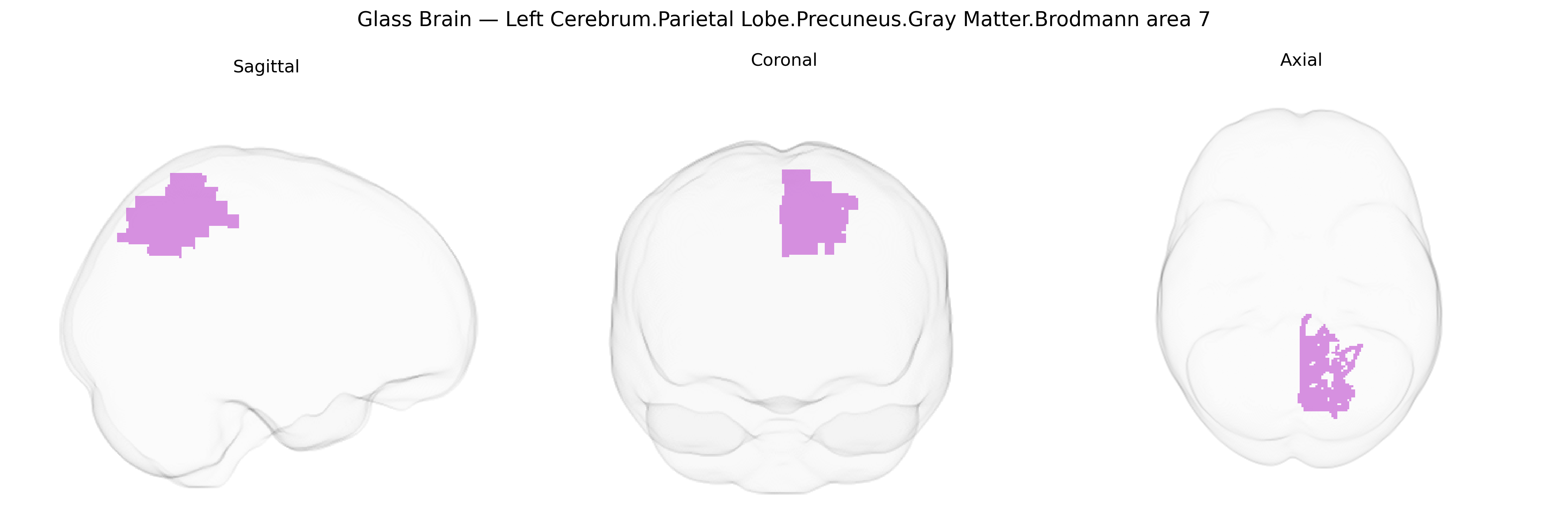
The bilateral Left Cerebrum.Parietal Lobe.Precuneus.Gray Matter.Brodmann area 7 corresponds to a region of superior posterior parietal cortex located in the medial parietal lobe, within the precuneus, and defined cytoarchitectonically as Brodmann area 7. This association cortex participates in visuospatial integration, coordination of visual and somatosensory information, and higher-order processes such as spatial attention, sensorimotor transformation, and aspects of imagery and self-referential processing. It forms part of large-scale networks connecting parietal, frontal, and occipital regions, contributing to visually guided actions, perception of spatial relationships, and integration of egocentric and allocentric reference frames. There is no direct link for this specific composite label; a related structure is the Precuneus.
The bilateral left precuneus (parietal lobe, Brodmann area 7) has been repeatedly implicated in imaging–genetics and GWAS studies of brain structure and function, although findings are typically reported for the precuneus or superior parietal cortex more generally rather than the specific Talairach BA7 label. Large-scale ENIGMA and UK Biobank analyses have identified common variants in and near genes such as HMGA2, IGF1, WNT3, KTN1, and microtubule/axon guidance genes that influence parietal and precuneus cortical thickness, surface area, and volume, with polygenic architecture overlapping cognitive performance, educational attainment, and intracranial volume. GWAS of intrinsic functional connectivity networks involving the default mode network, in which the precuneus is a central hub, implicate genes involved in synaptic function and myelination (e.g., MSRA, CNTNAP2-like loci) and show shared genetic variance with traits such as general intelligence and neuroticism. Disorder-focused imaging–genetic studies link risk loci for Alzheimer’s disease (e.g., APOE ε4 and CLU) and schizophrenia (e.g., CACNA1C, ZNF804A polygenic risk) to altered precuneus structure, hypometabolism, or connectivity, while autism spectrum disorder, attention-deficit/hyperactivity disorder, and major depression polygenic scores have been associated with default mode network/precuneus alterations. Overall, genetic influences on this region are highly polygenic and pleiotropic, contributing to individual differences in cognition, self-referential processing, and vulnerability to neuropsychiatric and neurodegenerative disorders.
Overview generated by GPT-4o (2026).
Region ID: 968
Hemisphere: bilateral
Atlas: Talairach labels 1mm
Full Quality Version: Download MP4
Full Quality Version: Download MP4
Wali Sidiqyar*, Gaurav Rudravaram*, Elyssa M. McMaster, Trent M. Schwartz, Adam M. Saunders, Kurt G. Schilling, Bennett A. Landman "Introducing SPINS: A Shared Public Visualization Library of Neuroanatomical Structures." Medical Imaging with Deep Learning- short paper
This resource is licensed under CC0 1.0 Universal (Public Domain).