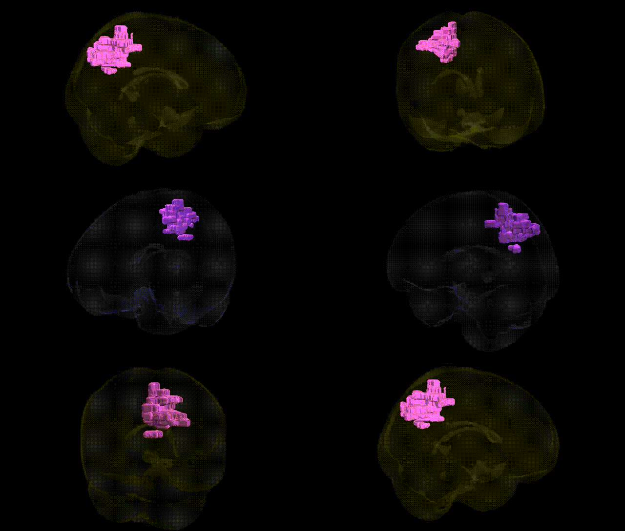
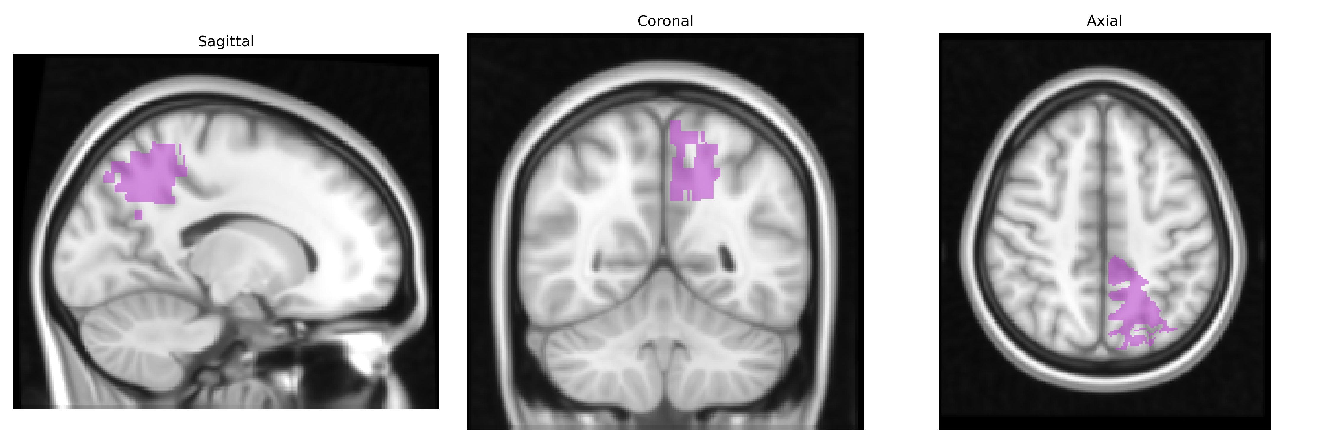
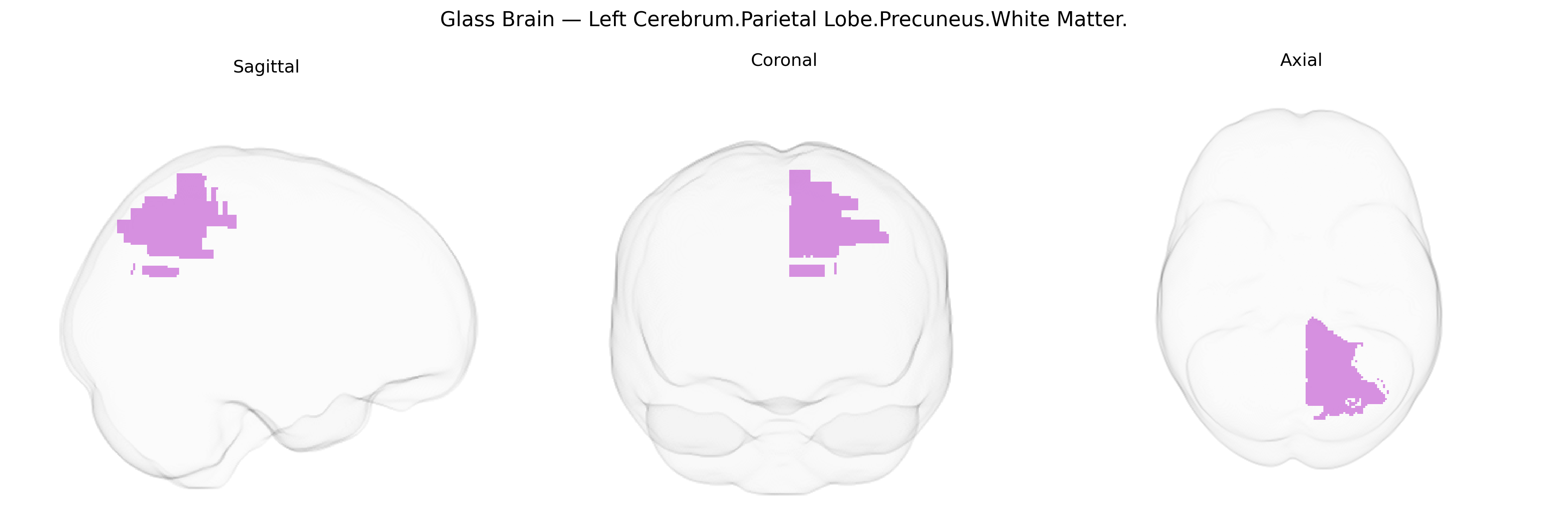
The bilateral Left Cerebrum.Parietal Lobe.Precuneus.White Matter corresponds to the intrahemispheric white matter tracts coursing beneath the cortical precuneus in the medial superior parietal lobule. This region contains long association fibers (such as components of the superior longitudinal fasciculus and cingulum bundle) and short U-fibers that interconnect adjacent and distant parietal, frontal, and occipital cortical areas, supporting visuospatial integration, self-referential processing, episodic memory, and aspects of attention and imagery. The white matter here serves as a major conduit linking the medial parietal cortex with default mode network nodes and other higher-order association cortices, and is supplied by penetrating branches of the posterior cerebral and, variably, the middle cerebral arterial systems. There is no direct Wikipedia article for the white matter of the precuneus; see the related cortical region: Precuneus.
The bilateral Left Cerebrum.Parietal Lobe.Precuneus.White Matter region, as defined in the Talairach 1 mm atlas, is consistently implicated in imaging genetics and GWAS of brain structure, cognition, and neuropsychiatric disorders, though findings are typically reported at the level of the broader precuneus or parietal white matter rather than this exact parcel. Large-scale neuroimaging GWAS (e.g., ENIGMA, UK Biobank) have identified common variants near genes such as HMGA2, IGF1, MAPT, and microtubule- or myelination-related genes that associate with precuneus or parietal lobe volume, cortical thickness, and white-matter microstructure (e.g., fractional anisotropy), suggesting roles for neurodevelopment, axonal growth, and myelin maintenance. Parietal and precuneus structural measures influenced by genetic variation have been linked to individual differences in general cognitive ability, memory, attention, and default-mode network connectivity. In psychiatric and neurological genetics, altered precuneus and adjacent parietal white-matter integrity shows genetic correlation with schizophrenia, major depression, bipolar disorder, autism spectrum disorder, and Alzheimer’s disease, often via polygenic risk scores that predict reduced volume or disrupted connectivity in this region. Several Alzheimer’s disease risk loci, including variants in APOE and CLU, show downstream associations with atrophy or hypometabolism in the precuneus and posterior cingulo-parietal hub, a key early site of pathology, while multiple sclerosis and other demyelinating or leukodystrophy-related genes contribute to lesion burden and microstructural damage in parietal white matter pathways that traverse or neighbor the precuneus.
Overview generated by GPT-4o (2026).
Region ID: 860
Hemisphere: bilateral
Atlas: Talairach labels 1mm
Full Quality Version: Download MP4
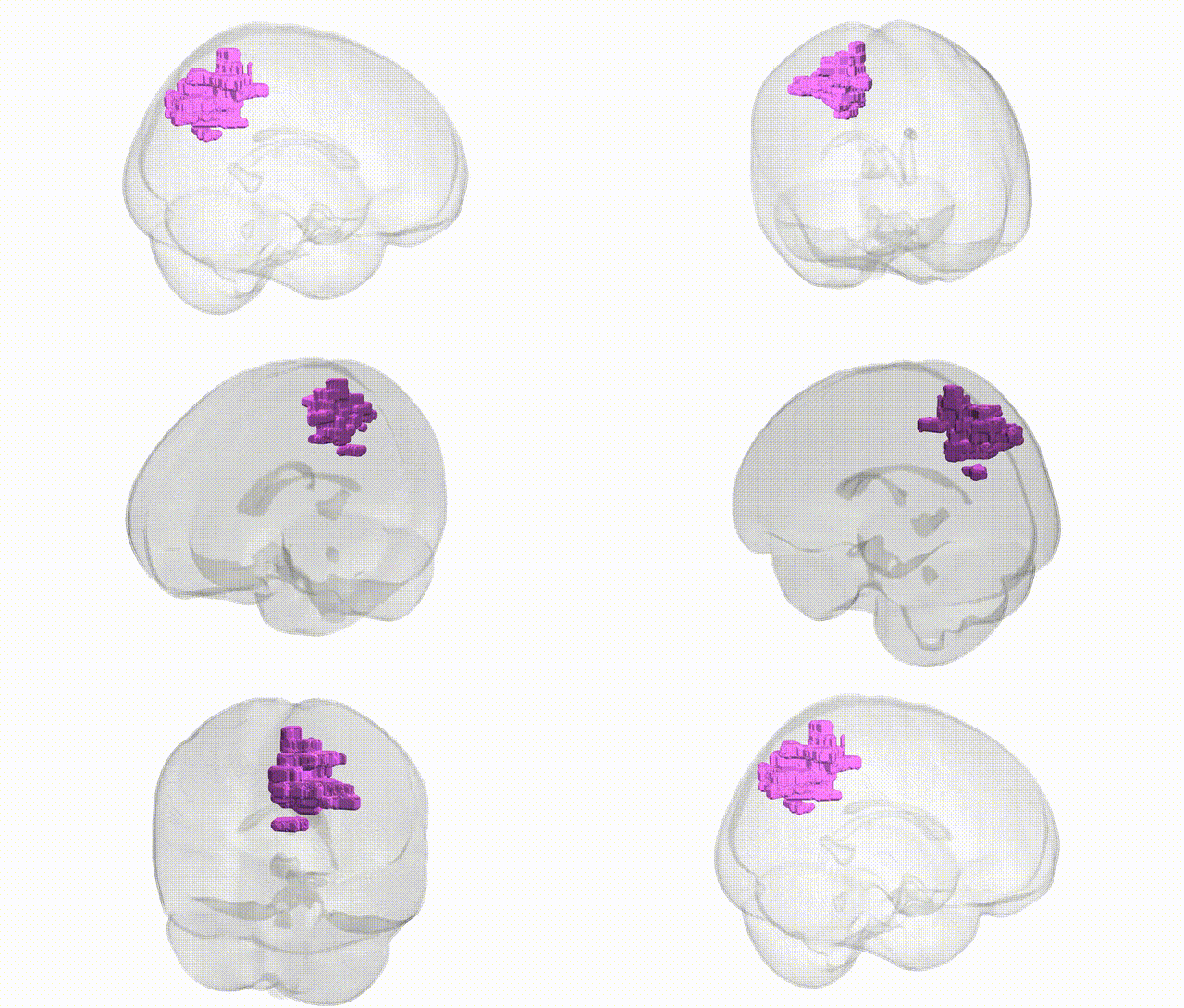
Full Quality Version: Download MP4
Wali Sidiqyar*, Gaurav Rudravaram*, Elyssa M. McMaster, Trent M. Schwartz, Adam M. Saunders, Kurt G. Schilling, Bennett A. Landman "Introducing SPINS: A Shared Public Visualization Library of Neuroanatomical Structures." Medical Imaging with Deep Learning- short paper
This resource is licensed under CC0 1.0 Universal (Public Domain).