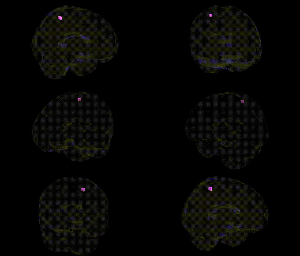
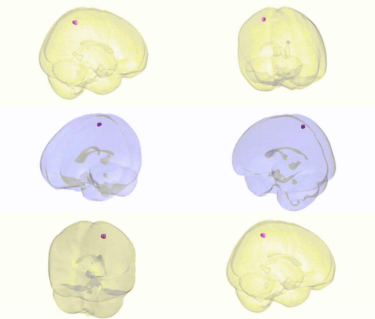
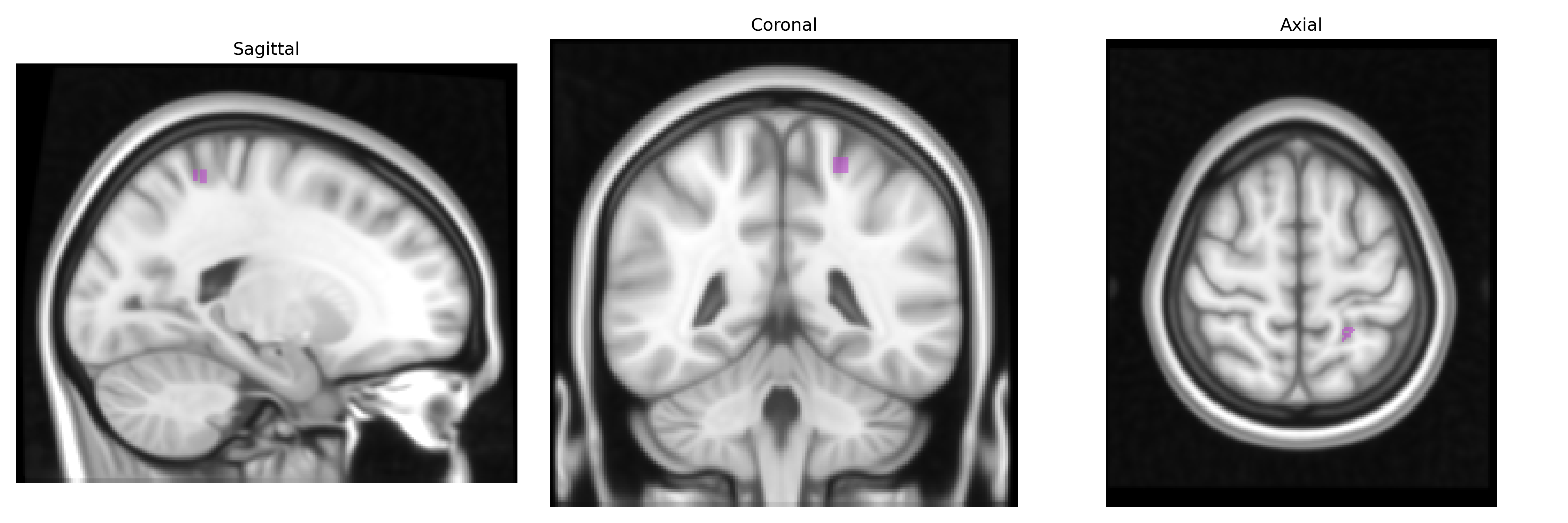
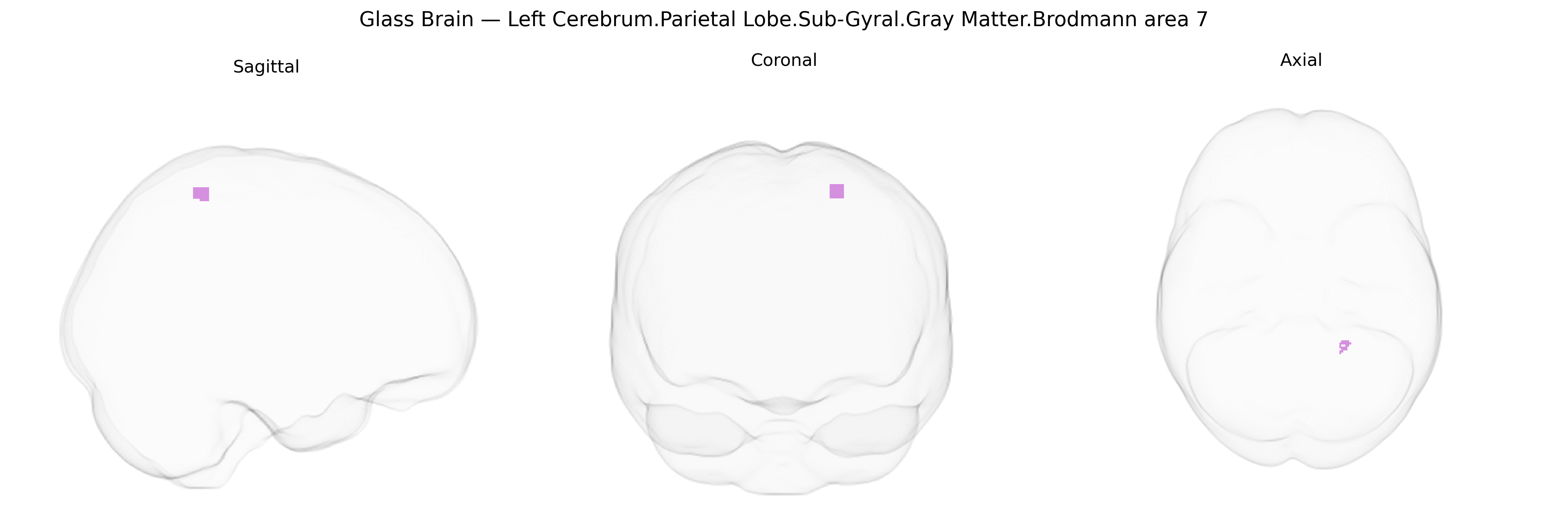
The bilateral Left Cerebrum.Parietal Lobe.Sub-Gyral.Gray Matter.Brodmann area 7 corresponds to a portion of the superior parietal lobule located in the posterior parietal cortex, deep to the cortical gyri, and composed primarily of association gray matter. Brodmann area 7 is involved in higher-order somatosensory processing, visuospatial integration, and sensorimotor coordination, contributing to functions such as spatial attention, body schema representation, and the guidance of visually directed movements. Neurons in this region integrate inputs from visual, somatosensory, and proprioceptive modalities, supporting the transformation of sensory information into coordinated actions and the perception of spatial relationships within the environment. Brodmann area 7
The bilateral left parietal sub-gyral gray matter of Brodmann area 7 (superior parietal lobule/precuneus region in the Talairach 1 mm atlas) has been implicated in multiple genetic and GWAS findings, largely via imaging-genetics and disorder-association studies that map variants onto cortical thickness, surface area, or activation in this territory. Polygenic influences related to neuronal development and synaptic function, including variants in pathways involving genes such as BDNF, NRG1, and glutamatergic signaling genes, have been associated with structural variance in superior parietal/BA7 cortex in large consortia (e.g., ENIGMA) and UK Biobank imaging GWAS, often in the context of global cortical measures. This region shows genetic correlations with intelligence, working memory, and visuospatial ability, consistent with traits linked to parietal structure and function; multiple GWAS of general cognitive ability identify loci (for example in genes like MAPT, MEF2C, and other neurodevelopmental genes) whose effects extend to parietal association cortices including BA7. Parietal BA7 is also commonly implicated in genetic risk for neurodevelopmental and psychiatric disorders—such as schizophrenia, autism spectrum disorder, and ADHD—where risk loci enriched in synaptic and neurodevelopmental pathways exhibit associations with altered parietal morphology or connectivity, and in neurodegenerative conditions like Alzheimer’s disease, where APOE and other GWAS-identified risk variants are linked to atrophy patterns involving the posterior parietal/precuneus region. Additionally, imaging-genetic work indicates that polygenic scores for disorders including schizophrenia, depression, and Alzheimer’s disease predict structural and functional variation in superior parietal/BA7 cortex, highlighting this area as a convergence zone where diverse genetic risks influence higher-order cognitive and visuospatial networks.
Overview generated by GPT-4o (2026).
Region ID: 1080
Hemisphere: bilateral
Atlas: Talairach labels 1mm
Full Quality Version: Download MP4
Full Quality Version: Download MP4
Wali Sidiqyar*, Gaurav Rudravaram*, Elyssa M. McMaster, Trent M. Schwartz, Adam M. Saunders, Kurt G. Schilling, Bennett A. Landman "Introducing SPINS: A Shared Public Visualization Library of Neuroanatomical Structures." Medical Imaging with Deep Learning- short paper
This resource is licensed under CC0 1.0 Universal (Public Domain).