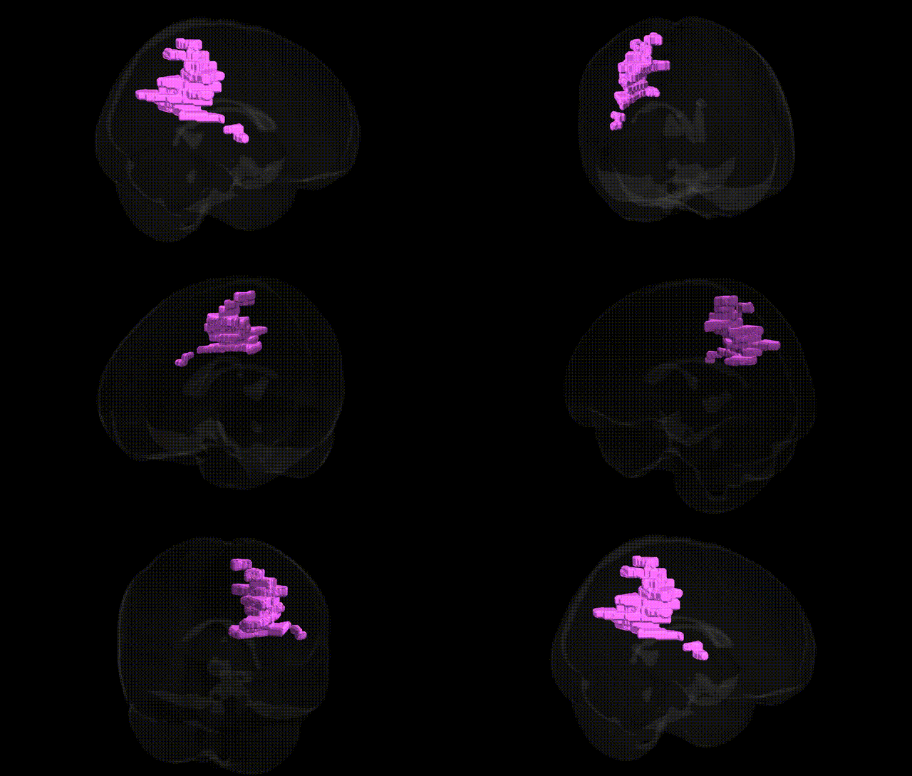
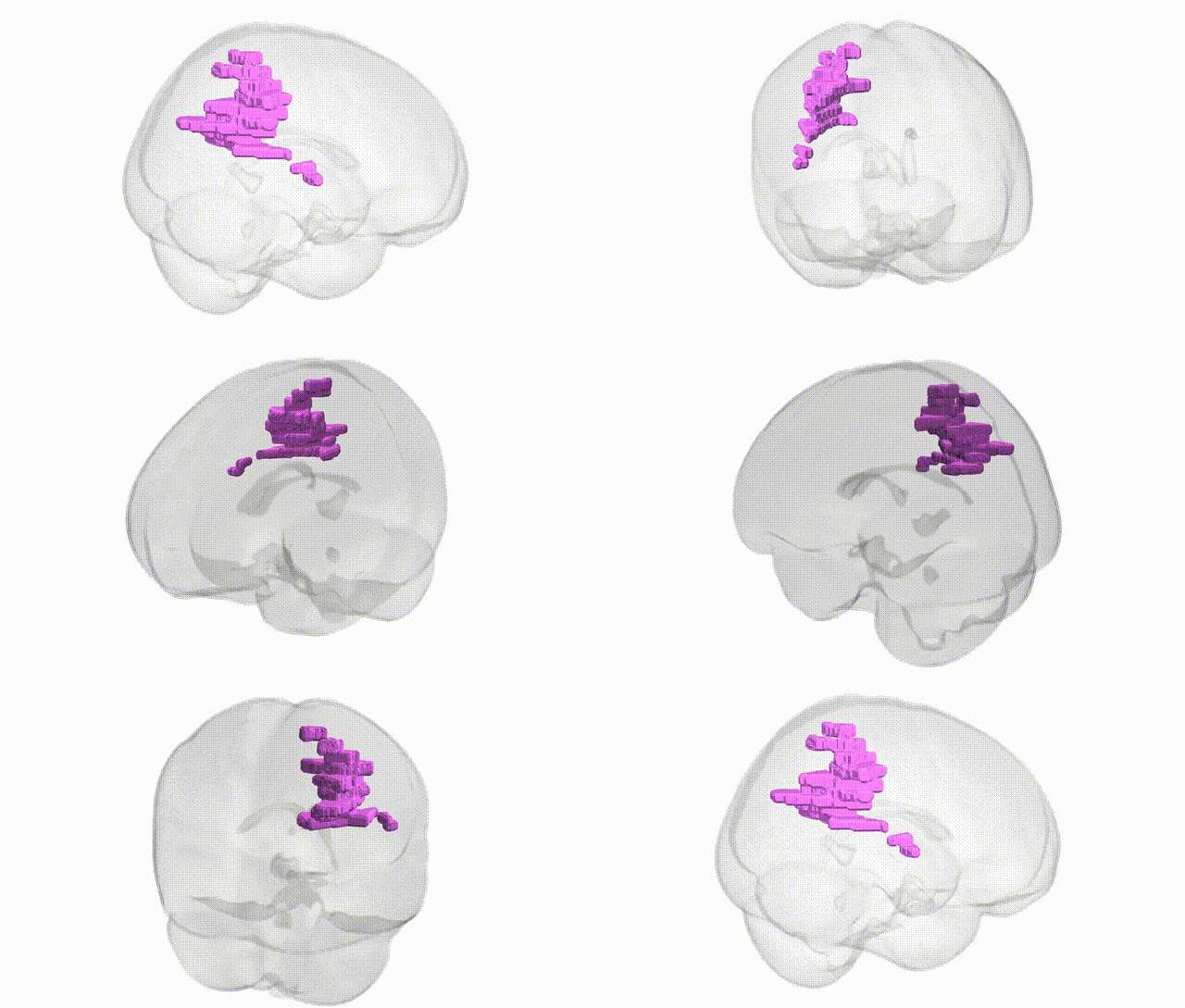
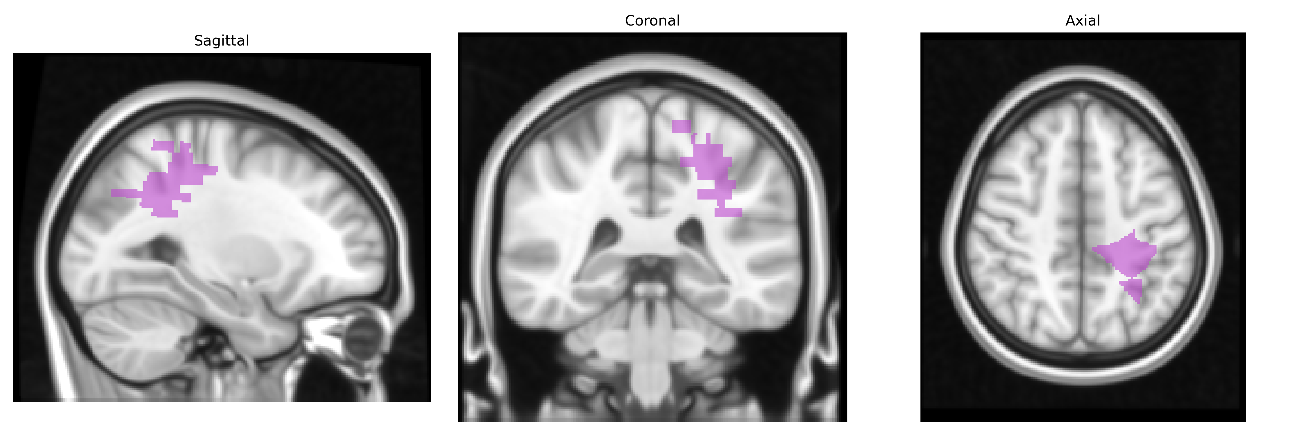
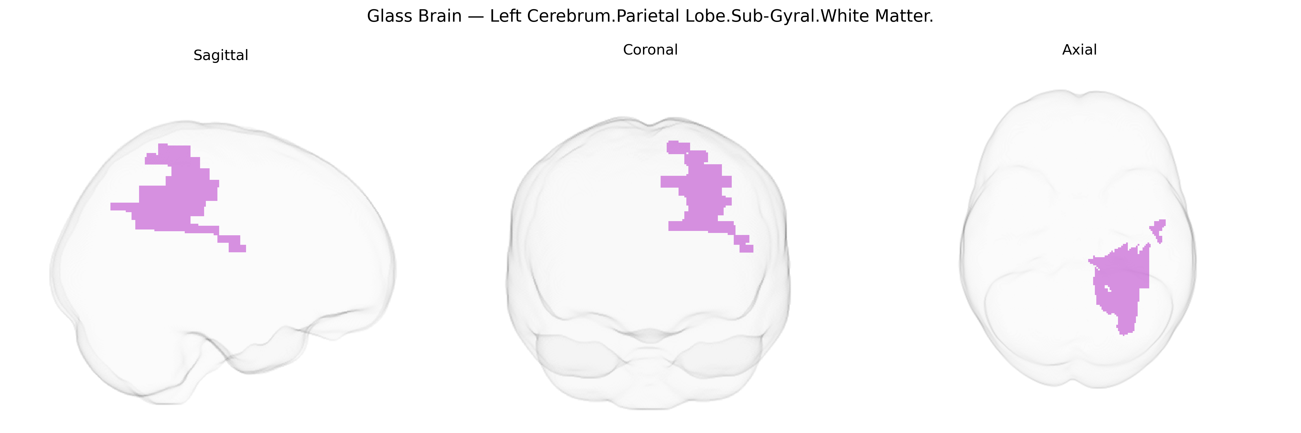
The bilateral Left Cerebrum.Parietal Lobe.Sub-Gyral.White Matter refers to the deep white matter lying beneath the cortical gyri of the left parietal lobe. This region consists primarily of myelinated axonal fibers that interconnect parietal cortical areas with other cortical and subcortical structures, supporting integration of somatosensory information, visuospatial processing, attention, and aspects of language and numerical cognition. As part of the parietal white matter, it contributes to major association pathways such as segments of the superior longitudinal fasciculus and other intrahemispheric tracts that enable communication between frontal, temporal, and occipital lobes. There is no direct Wikipedia article for this precise sub-gyral white matter label; a closely related structure is the Parietal lobe.
Genetic associations involving the bilateral Left Cerebrum.Parietal Lobe.Sub-Gyral.White Matter, as defined in the Talairach 1 mm atlas, derive largely from GWAS and imaging-genetics studies of parietal lobe volume, cortical thickness, and white matter microstructure (e.g., fractional anisotropy), as well as from clinical genetics of neurodevelopmental and neuropsychiatric disorders affecting parietal circuits. Genome-wide analyses of parietal lobe structure have implicated loci in or near genes involved in neurodevelopment, axonal guidance, and myelination, including variants in pathways such as WNT signaling, cell adhesion (e.g., cadherins, contactins), and oligodendrocyte function; several large consortia (e.g., ENIGMA and UK Biobank–based studies) report polygenic influences spread across the genome rather than single large-effect variants, with modest but consistent heritability for parietal white matter metrics. Clinically, copy-number variants and rare mutations in genes associated with neurodevelopmental disorders (including autism spectrum disorder, intellectual disability, and specific learning disorders such as developmental dyscalculia), as well as genes implicated in schizophrenia and attention-deficit/hyperactivity disorder, have been linked to altered parietal structure and connectivity, encompassing sub-gyral white matter pathways that integrate parietal with frontal and temporal regions. Additional associations arise from GWAS of cognitive traits such as working memory, visuospatial processing, and mathematical ability, where risk alleles correlate with differences in parietal lobe morphology or white matter integrity; vascular and demyelinating disorders (e.g., cerebral small vessel disease and multiple sclerosis) show genetic risk architectures (including variants in immune and endothelial genes) that are expressed, in part, through lesion burden and microstructural change in parietal white matter. Overall, current evidence indicates that the sub-gyral parietal white matter is influenced by a highly polygenic architecture in which common and rare variants impacting neurodevelopment, myelination, and connectivity contribute to individual differences in structure and to risk for cognitive and psychiatric phenotypes, although gene–region relationships remain probabilistic and spatially coarse rather than specific to this exact Talairach-defined parcel.
Overview generated by GPT-4o (2026).
Region ID: 796
Hemisphere: bilateral
Atlas: Talairach labels 1mm
Full Quality Version: Download MP4
Full Quality Version: Download MP4
Wali Sidiqyar*, Gaurav Rudravaram*, Elyssa M. McMaster, Trent M. Schwartz, Adam M. Saunders, Kurt G. Schilling, Bennett A. Landman "Introducing SPINS: A Shared Public Visualization Library of Neuroanatomical Structures." Medical Imaging with Deep Learning- short paper
This resource is licensed under CC0 1.0 Universal (Public Domain).