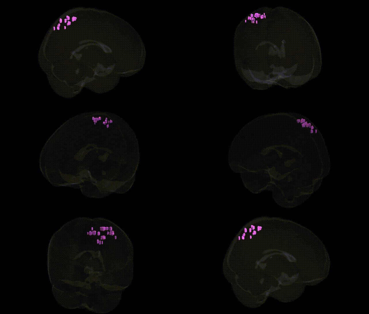
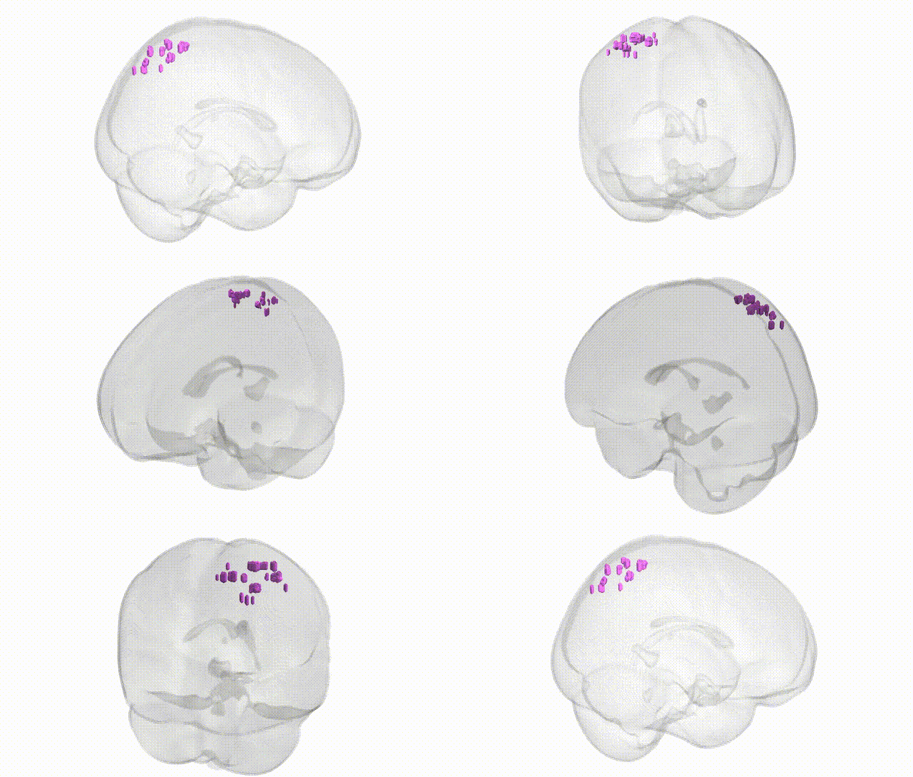
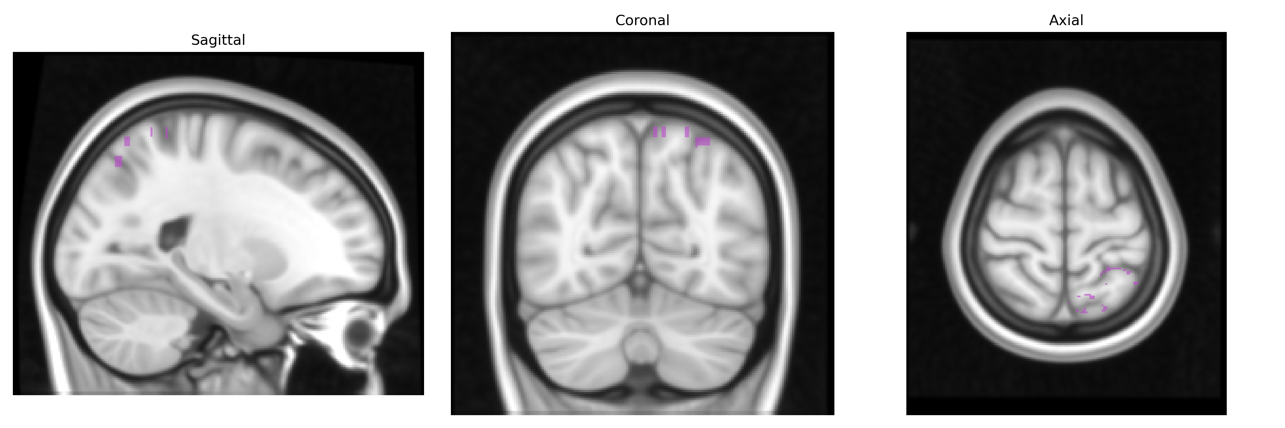
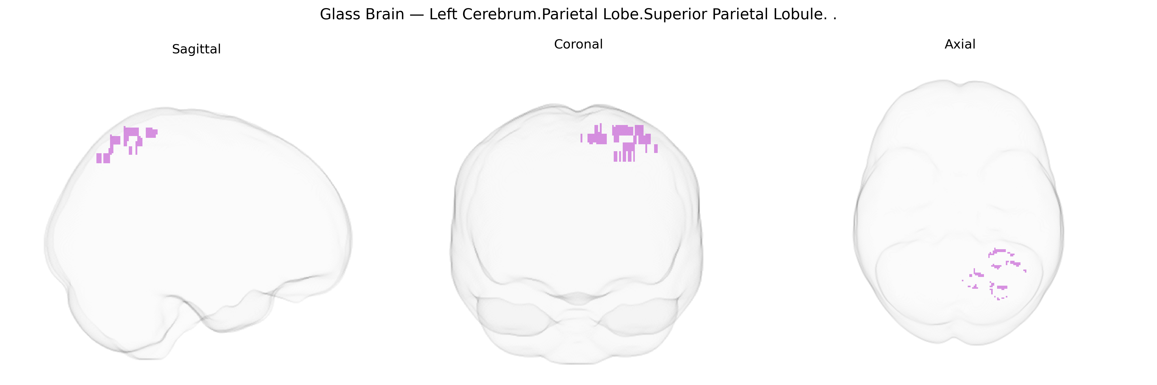
The bilateral Left Cerebrum.Parietal Lobe.Superior Parietal Lobule corresponds to the superior parietal lobule (SPL), a dorsal parietal cortical region situated posterior to the postcentral gyrus and superior to the intraparietal sulcus, extending toward the parieto‑occipital boundary. Cytoarchitectonically, it includes parts of Brodmann areas 5 and 7. The SPL is critically involved in multimodal integration of somatosensory, visual, and proprioceptive information, supporting spatial perception, visuomotor coordination, body‑schema representation, and aspects of attention and working memory related to spatial processing. It contributes to the guidance of reaching and grasping movements and to the transformation of sensory coordinates into action-oriented representations. Lesions in this region can result in deficits such as optic ataxia, impaired spatial orientation, and disturbances in sensorimotor integration. Superior parietal lobule
The bilateral superior parietal lobule (SPL) of the left cerebrum, as defined in the Talairach 1 mm atlas, has been implicated in multiple genetic and genome-wide association studies, primarily through its roles in visuospatial processing, attention, working memory, and sensorimotor integration. Polygenic influences on SPL structure (cortical thickness, surface area, and gyrification) have been identified in large-scale imaging-genetics consortia such as ENIGMA and UK Biobank, with heritable variation linked broadly to neurodevelopmental and synaptic genes, although individual SNP effects are typically small. GWAS and related analyses have associated SPL measures or activation patterns with cognitive performance traits (e.g., general intelligence, working memory, mathematical ability), attention and visuospatial skills, and motor coordination, often implicating genes involved in axon guidance, neuronal migration, and plasticity (for example, pathways including NRGN, BDNF-related signaling, and other synaptic scaffolding genes, though not uniquely restricted to SPL). Structural and functional alterations in the superior parietal region show genetic correlations with neurodevelopmental and psychiatric disorders such as schizophrenia, autism spectrum disorder, attention-deficit/hyperactivity disorder, and major depressive disorder, largely via polygenic risk scores that predict parietal morphology and connectivity. Neurodegenerative and white-matter–related loci (e.g., APOE and other Alzheimer’s disease risk variants) have been associated with atrophy or hypometabolism in parietal areas, including SPL, in aging and dementia cohorts. Overall, genetic findings support the SPL as a polygenically influenced hub whose structure and function mediate the effects of widely distributed neurocognitive and neuropsychiatric risk variants rather than being driven by a small set of region-specific genes.
Overview generated by GPT-4o (2026).
Region ID: 1037
Hemisphere: bilateral
Atlas: Talairach labels 1mm
Full Quality Version: Download MP4
Full Quality Version: Download MP4
Wali Sidiqyar*, Gaurav Rudravaram*, Elyssa M. McMaster, Trent M. Schwartz, Adam M. Saunders, Kurt G. Schilling, Bennett A. Landman "Introducing SPINS: A Shared Public Visualization Library of Neuroanatomical Structures." Medical Imaging with Deep Learning- short paper
This resource is licensed under CC0 1.0 Universal (Public Domain).