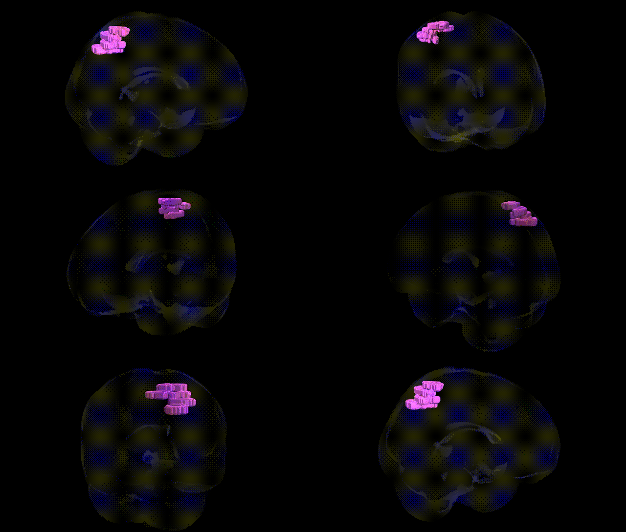
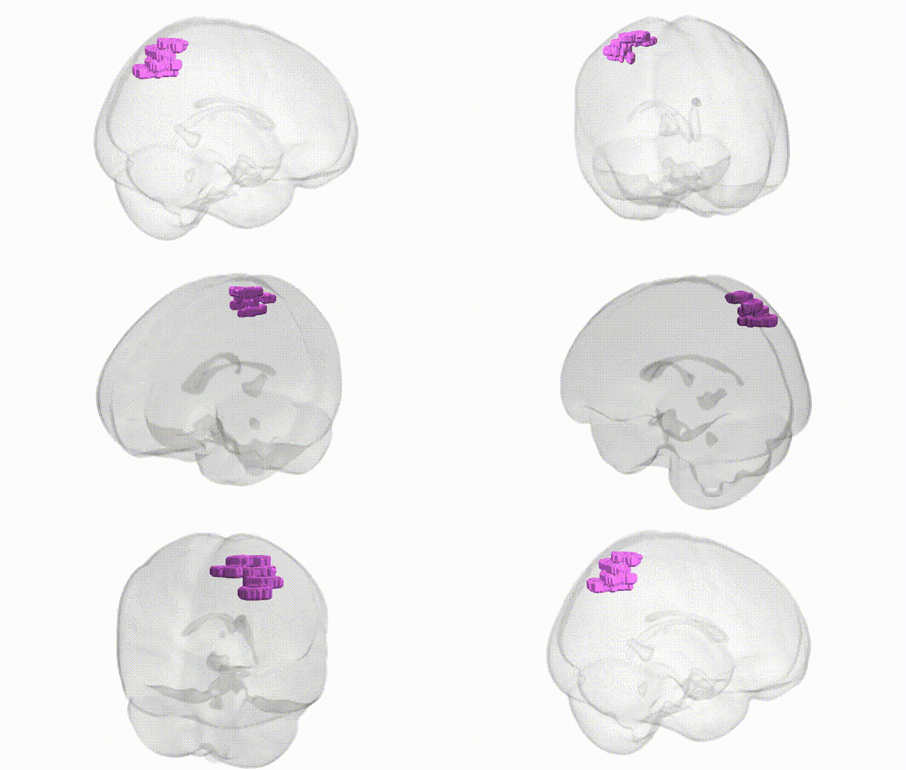
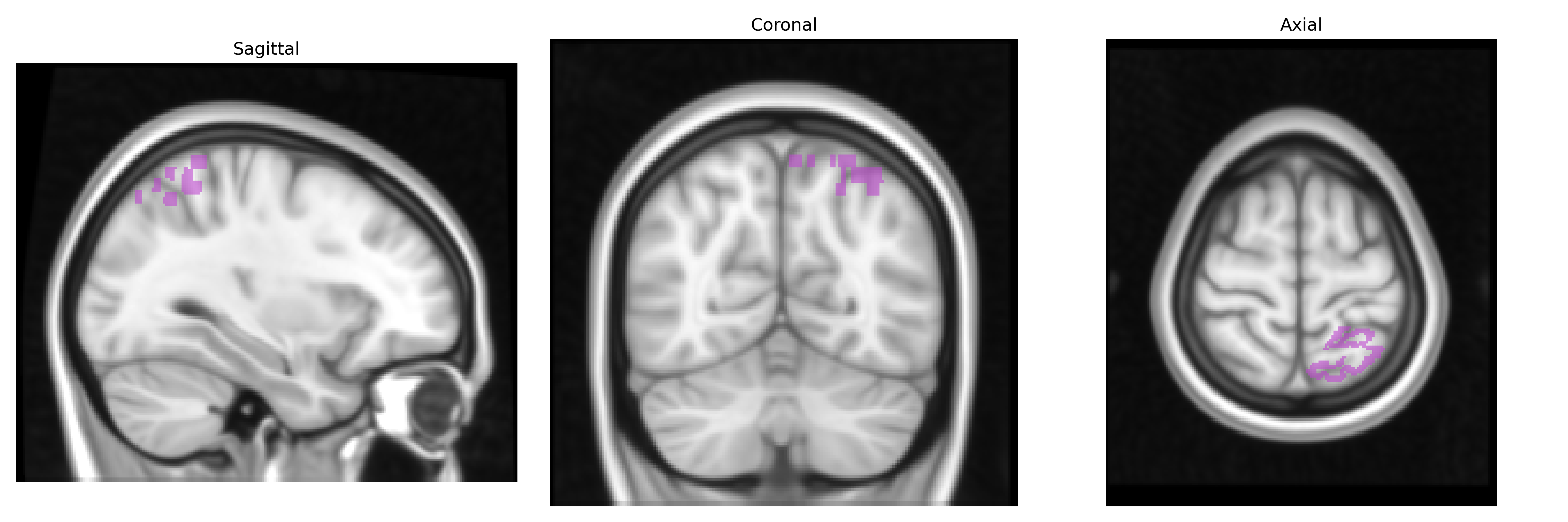
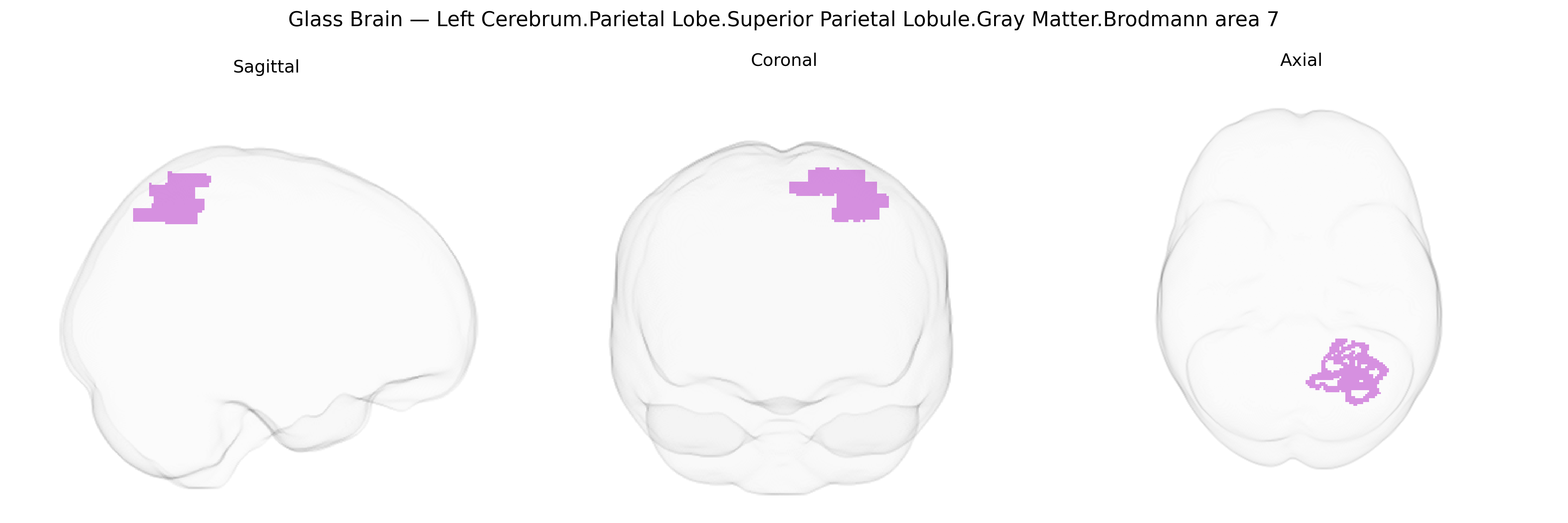
The bilateral Left Cerebrum.Parietal Lobe.Superior Parietal Lobule.Gray Matter.Brodmann area 7 corresponds to an association cortex region in the superior parietal lobule involved in higher-order somatosensory integration, visuospatial processing, and sensorimotor coordination. Cytoarchitectonically, Brodmann area 7 is characterized by a granular isocortical structure with prominent layer IV and well-developed supragranular layers that support complex multimodal integration. Functionally, this region contributes to spatial attention, visuomotor transformations (such as guiding reaching and grasping), and the internal representation of body schema and extrapersonal space. Through extensive reciprocal connections with primary and secondary somatosensory cortices, visual association areas, and frontal motor-related regions, Brodmann area 7 plays a crucial role in transforming sensory input into coordinated action and in constructing coherent spatial representations of the environment and the body. Brodmann area 7
The bilateral superior parietal lobule (SPL; Brodmann area 7) has been implicated in multiple genetic and GWAS-derived associations, primarily through imaging genetics and large-scale neuroimaging consortia such as ENIGMA and UK Biobank. Heritability estimates for gray matter volume and cortical surface area in BA7 are moderate to high, with polygenic influences distributed across many loci rather than a few large-effect variants. Common variants in genes involved in neurodevelopment, synaptic plasticity, and axon guidance (for example, BDNF, NRG1, and several calcium-channel and glutamatergic signaling genes) have been repeatedly associated with parietal cortical thickness, surface area, or activation patterns in working memory and visuospatial tasks that rely on SPL function. GWAS meta-analyses of cortical structure have identified loci near genes such as HMGA2, IGF1, and microtubule- or cytoskeleton-related genes that contribute to interindividual differences in parietal lobe morphology, including superior parietal gray matter. In psychiatric genetics, risk variants for schizophrenia, bipolar disorder, autism spectrum disorder, and ADHD show downstream effects on SPL structure and connectivity, with case–control and polygenic risk score studies linking higher genetic liability for these conditions to reduced gray matter volume, altered cortical thickness, or atypical functional connectivity in BA7. Neurodegenerative conditions such as Alzheimer’s disease and posterior cortical atrophy also exhibit genetically modulated vulnerability in the superior parietal lobule, where APOE ε4 and other AD-related loci are associated with accelerated atrophy or hypometabolism in parietal regions. Additionally, GWAS of cognitive traits (general intelligence, mathematical ability, and visuospatial reasoning) and sensorimotor functions have reported overlapping polygenic architectures that influence both performance and SPL structure or activation, underscoring this region’s role as a genetically sensitive hub for higher-order visuospatial, attention, and integration processes.
Overview generated by GPT-4o (2026).
Region ID: 1039
Hemisphere: bilateral
Atlas: Talairach labels 1mm
Full Quality Version: Download MP4
Full Quality Version: Download MP4
Wali Sidiqyar*, Gaurav Rudravaram*, Elyssa M. McMaster, Trent M. Schwartz, Adam M. Saunders, Kurt G. Schilling, Bennett A. Landman "Introducing SPINS: A Shared Public Visualization Library of Neuroanatomical Structures." Medical Imaging with Deep Learning- short paper
This resource is licensed under CC0 1.0 Universal (Public Domain).