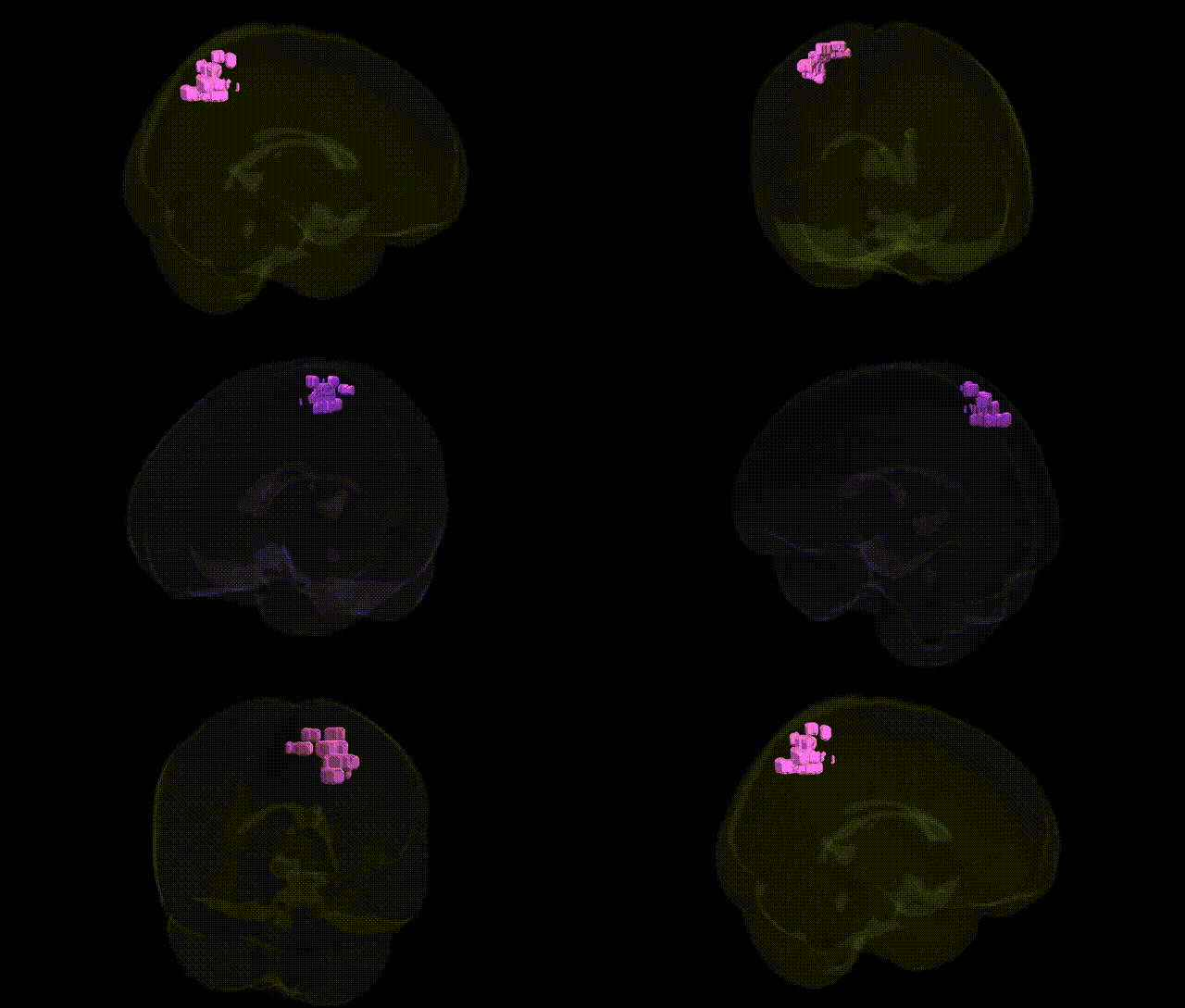
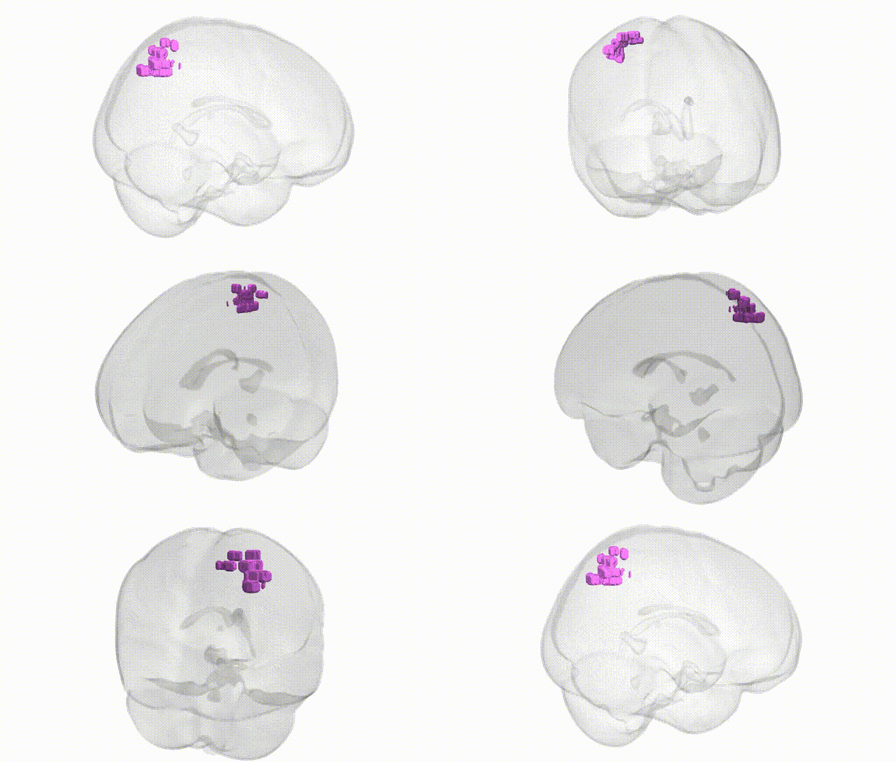
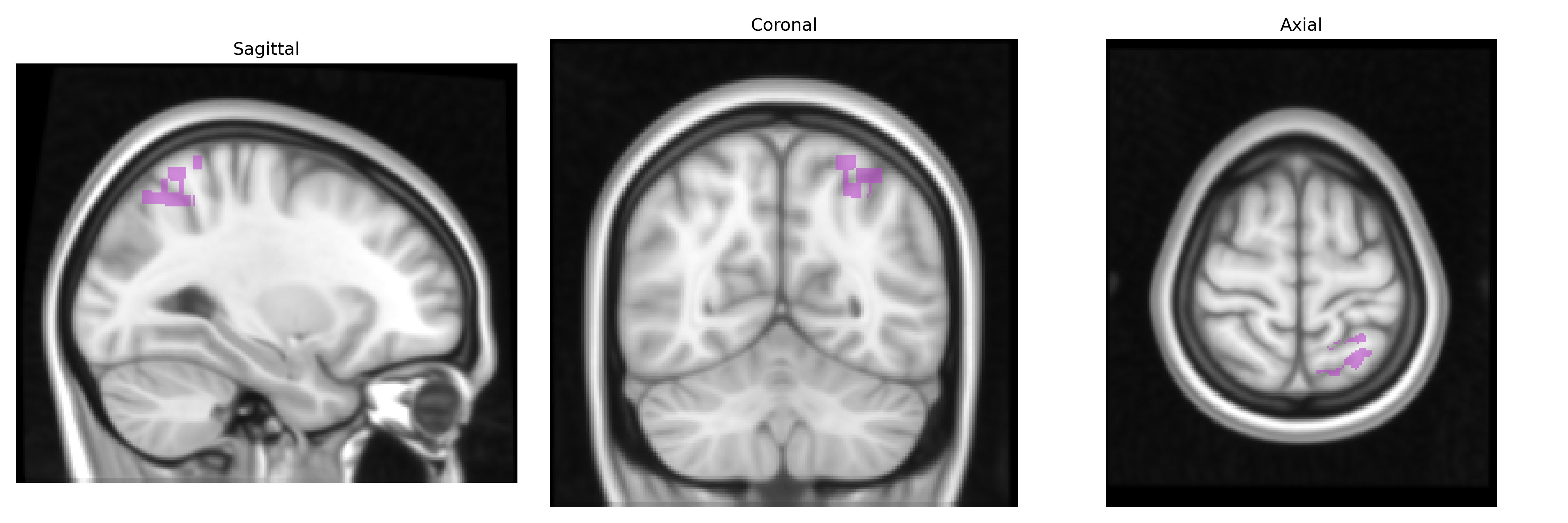
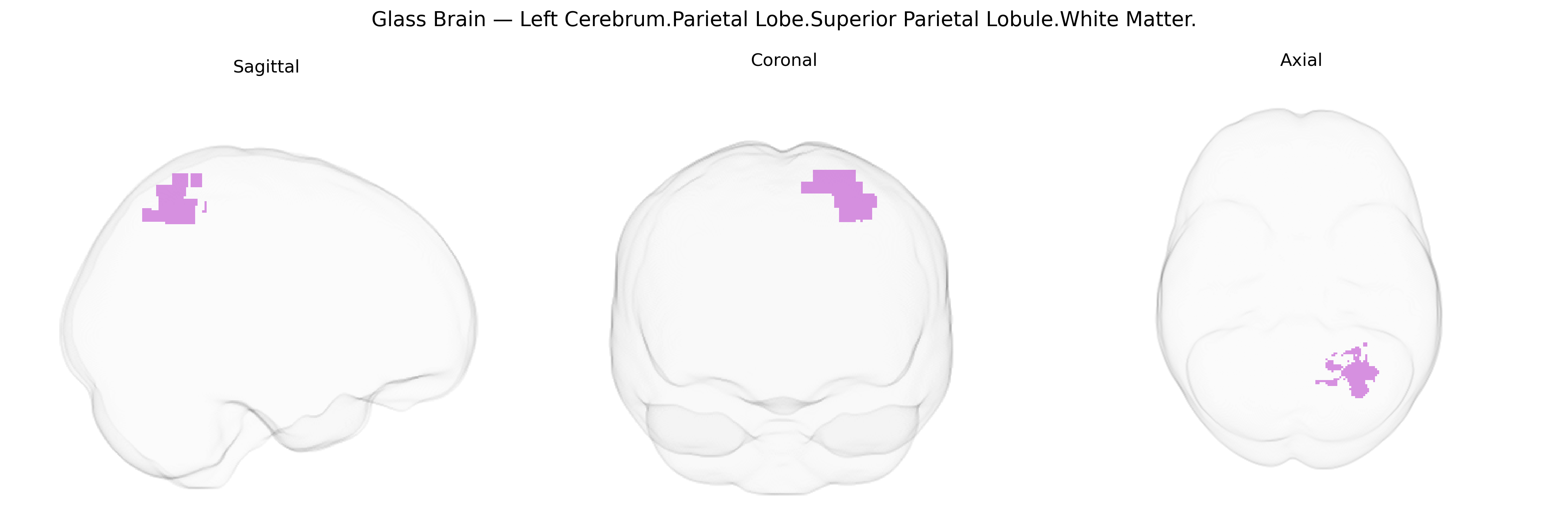
The bilateral Left Cerebrum.Parietal Lobe.Superior Parietal Lobule.White Matter corresponds to the deep myelinated fiber tracts underlying the superior parietal lobule in the left hemisphere, as defined in the Talairach 1 mm atlas. These white matter pathways connect superior parietal cortical regions—with roles in spatial perception, sensorimotor integration, and visuomotor coordination—to other parietal, frontal, and occipital areas, supporting networks involved in attention, body schema, and visually guided movement. Fibers in this region include components of major association bundles such as segments of the superior longitudinal fasciculus, which link parietal association cortex with premotor and prefrontal areas, thereby facilitating integration of somatosensory and visual information for higher-order cognitive and motor functions. There is no direct link for this exact white matter label; a related cortical region is the Superior parietal lobule.
The bilateral Left Cerebrum.Parietal Lobe.Superior Parietal Lobule.White Matter (as defined in the Talairach 1 mm atlas) has been implicated in several genetic and GWAS-based associations, largely via imaging-genetics and neurodevelopmental studies of parietal white-matter integrity and cortical organization. Variants in genes regulating axonal guidance and myelination (for example, NTRK1/2, ROBO1/2, L1CAM, and oligodendrocyte-related genes such as MAG and MBP) have been associated with microstructural properties of superior parietal white-matter tracts measured by diffusion MRI, often in or near the superior longitudinal fasciculus and related parietal pathways. Large-scale GWAS of brain structure (e.g., ENIGMA and UK Biobank) have reported loci influencing parietal cortical thickness and surface area, with effects in genes involved in neurodevelopment (such as MIR137, HMGA2, and WNT pathway genes) that likely extend to adjacent white matter. Clinically, genetic risk for neurodevelopmental and psychiatric conditions—including autism spectrum disorder, schizophrenia, and ADHD—has been linked to altered superior parietal white-matter connectivity, with polygenic risk scores for these disorders correlating with parietal lobe microstructure in imaging-genetics cohorts. In addition, common variants associated with general cognitive ability, visuospatial processing, and educational attainment show anatomical correlates in parietal regions, suggesting that genetic influences on cognition partly act through development and maintenance of superior parietal white matter.
Overview generated by GPT-4o (2026).
Region ID: 1041
Hemisphere: bilateral
Atlas: Talairach labels 1mm
Full Quality Version: Download MP4
Full Quality Version: Download MP4
Wali Sidiqyar*, Gaurav Rudravaram*, Elyssa M. McMaster, Trent M. Schwartz, Adam M. Saunders, Kurt G. Schilling, Bennett A. Landman "Introducing SPINS: A Shared Public Visualization Library of Neuroanatomical Structures." Medical Imaging with Deep Learning- short paper
This resource is licensed under CC0 1.0 Universal (Public Domain).