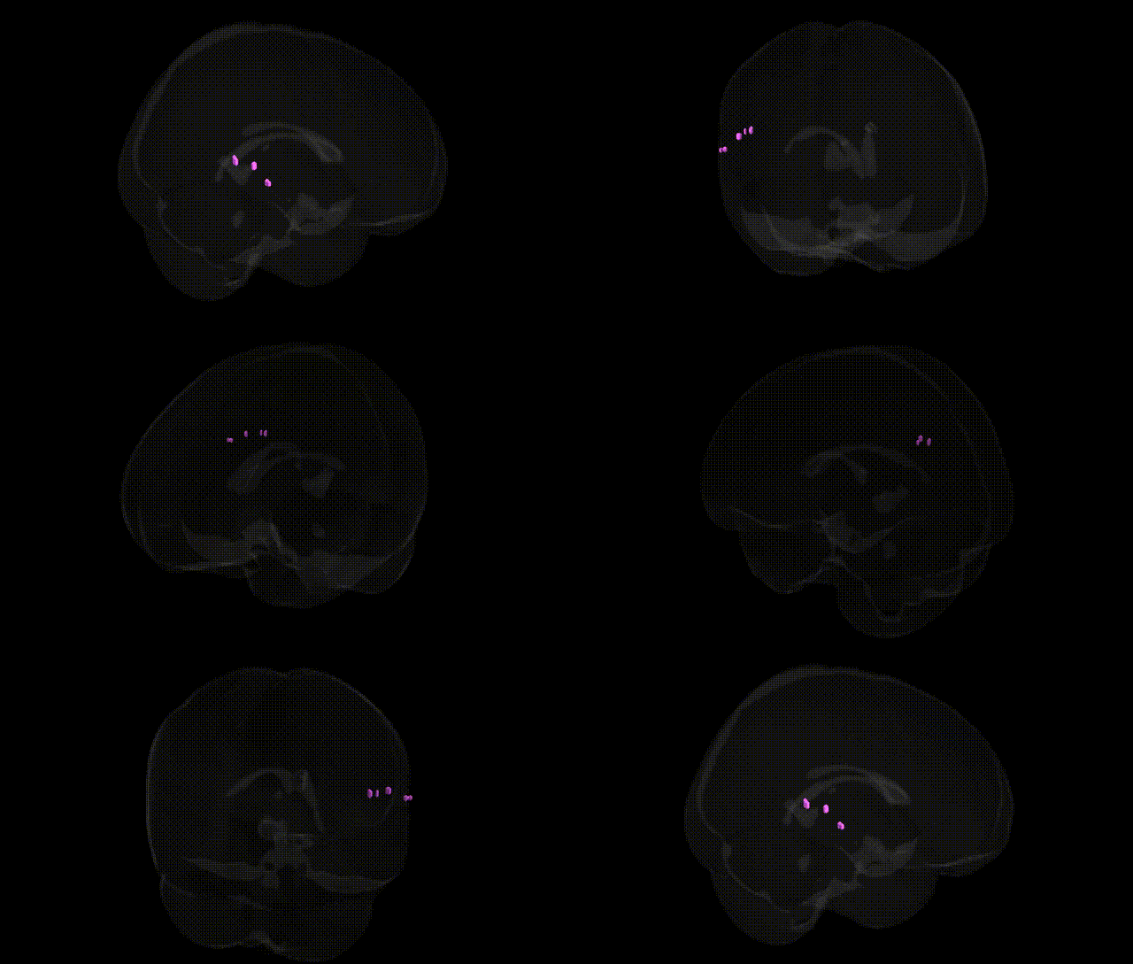
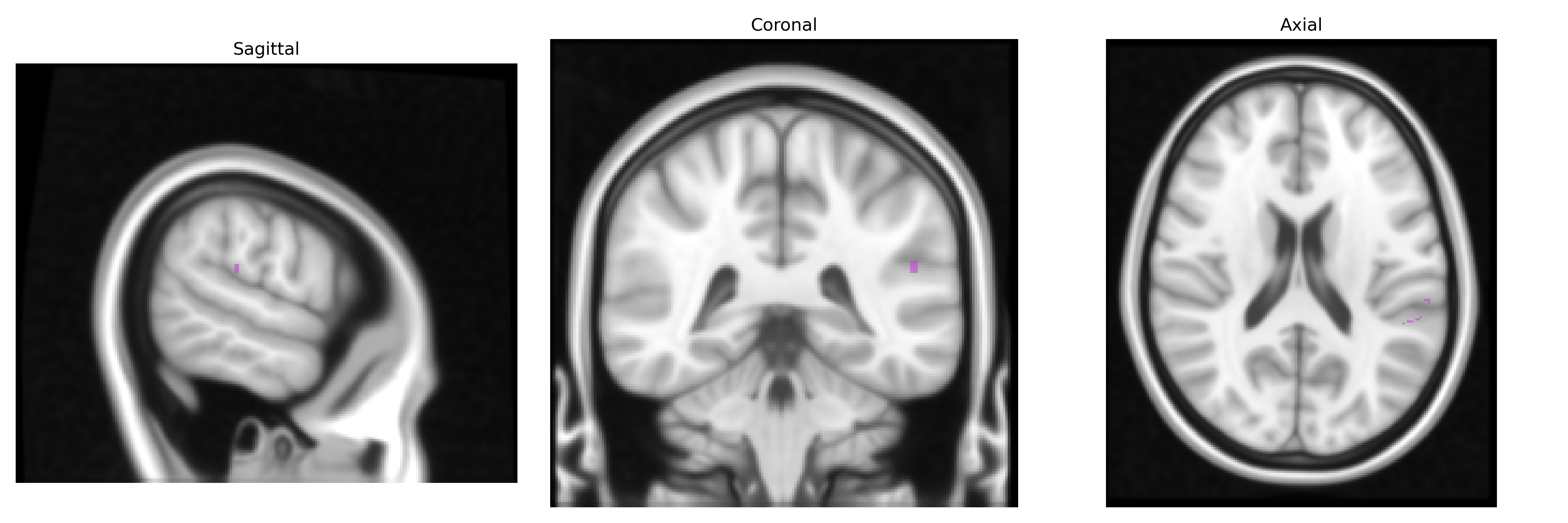
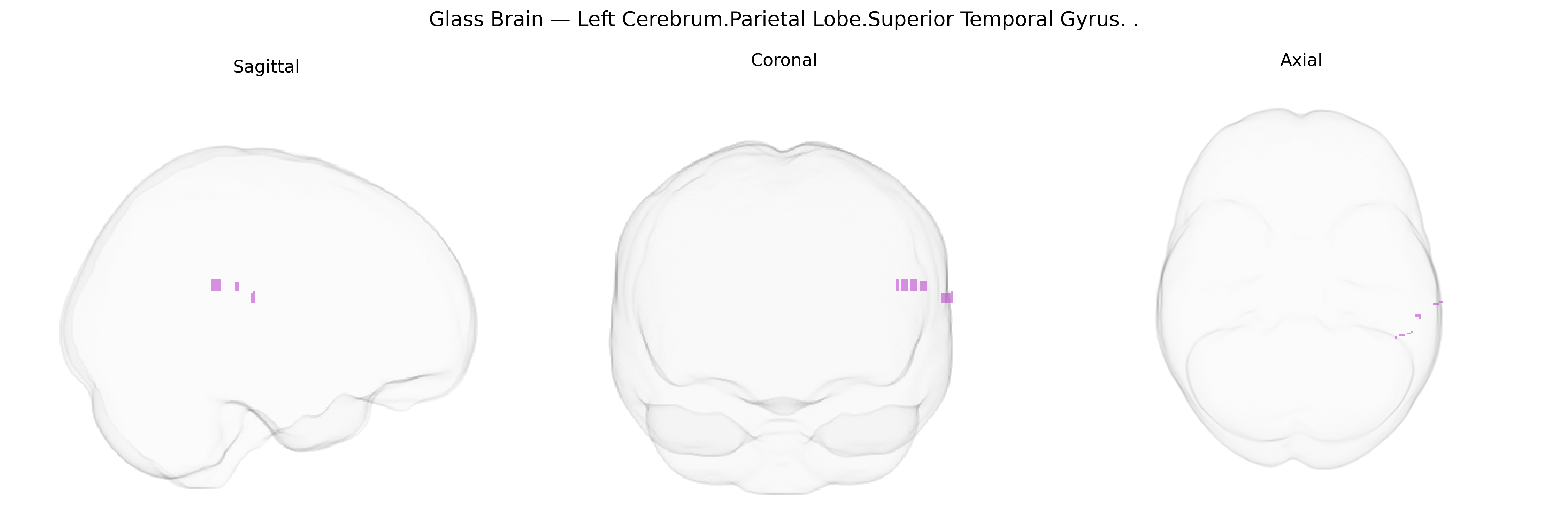
The bilateral Left Cerebrum.Parietal Lobe.Superior Temporal Gyrus label in the Talairach 1 mm atlas corresponds primarily to the superior temporal gyrus (STG) of the left hemisphere, a cortical region along the lateral surface of the temporal lobe bordering the lateral sulcus and functionally interacting with adjacent parietal areas at the temporoparietal junction. This region includes key components of the auditory cortex and language-related areas (notably parts of Wernicke’s area), supporting complex auditory processing, phonological decoding, speech comprehension, and aspects of semantic integration, while also contributing to multimodal sensory integration and social-cognitive functions such as processing of prosody and biological motion. The STG is highly interconnected with frontal, parietal, and limbic regions, and its structural and functional integrity is critical in disorders involving language, auditory perception (including hallucinations), and social communication. Superior temporal gyrus
The bilateral superior temporal gyrus (STG) in the parietal/temporal region has been repeatedly implicated in genetic studies of language, social cognition, and psychiatric risk, with GWAS and imaging–genetics work showing that common variants influence its structure and function. Variants in FOXP2, CNTNAP2, DCDC2, KIAA0319, and ROBO1 have been associated with STG morphology and activation patterns in language tasks, and polygenic scores for educational attainment and cognitive ability correlate with surface area and cortical thickness of temporal–parietal regions that include the STG. Large-scale neuroimaging GWAS from consortia such as ENIGMA and UK Biobank have identified multiple loci (e.g., in MIR124-1, HMGA2, and genes involved in neurodevelopmental pathways) associated with temporal lobe cortical thickness and surface area, including the superior temporal gyrus, and these same loci often show pleiotropic associations with schizophrenia, bipolar disorder, autism spectrum disorder, and major depressive disorder. Schizophrenia risk variants in genes such as CACNA1C, GRM3, and ZNF804A have been linked to altered superior temporal activation during auditory and language processing, while structural imaging GWAS indicate that polygenic risk for schizophrenia and autism relates to reduced STG volume and atypical asymmetry. In addition, GWAS of tinnitus, auditory hallucinations, and musical aptitude highlight temporal–parietal regions including the STG, and genetic variation in serotonin, glutamate, and GABA-related genes has been tied to functional differences in this area in disorders such as social anxiety and autism, underscoring the region’s role as a convergence zone for genetic influences on language, perception, and social–emotional processing.
Overview generated by GPT-4o (2026).
Region ID: 783
Hemisphere: bilateral
Atlas: Talairach labels 1mm
Full Quality Version: Download MP4
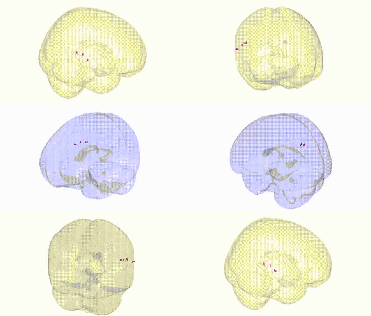
Full Quality Version: Download MP4
Wali Sidiqyar*, Gaurav Rudravaram*, Elyssa M. McMaster, Trent M. Schwartz, Adam M. Saunders, Kurt G. Schilling, Bennett A. Landman "Introducing SPINS: A Shared Public Visualization Library of Neuroanatomical Structures." Medical Imaging with Deep Learning- short paper
This resource is licensed under CC0 1.0 Universal (Public Domain).