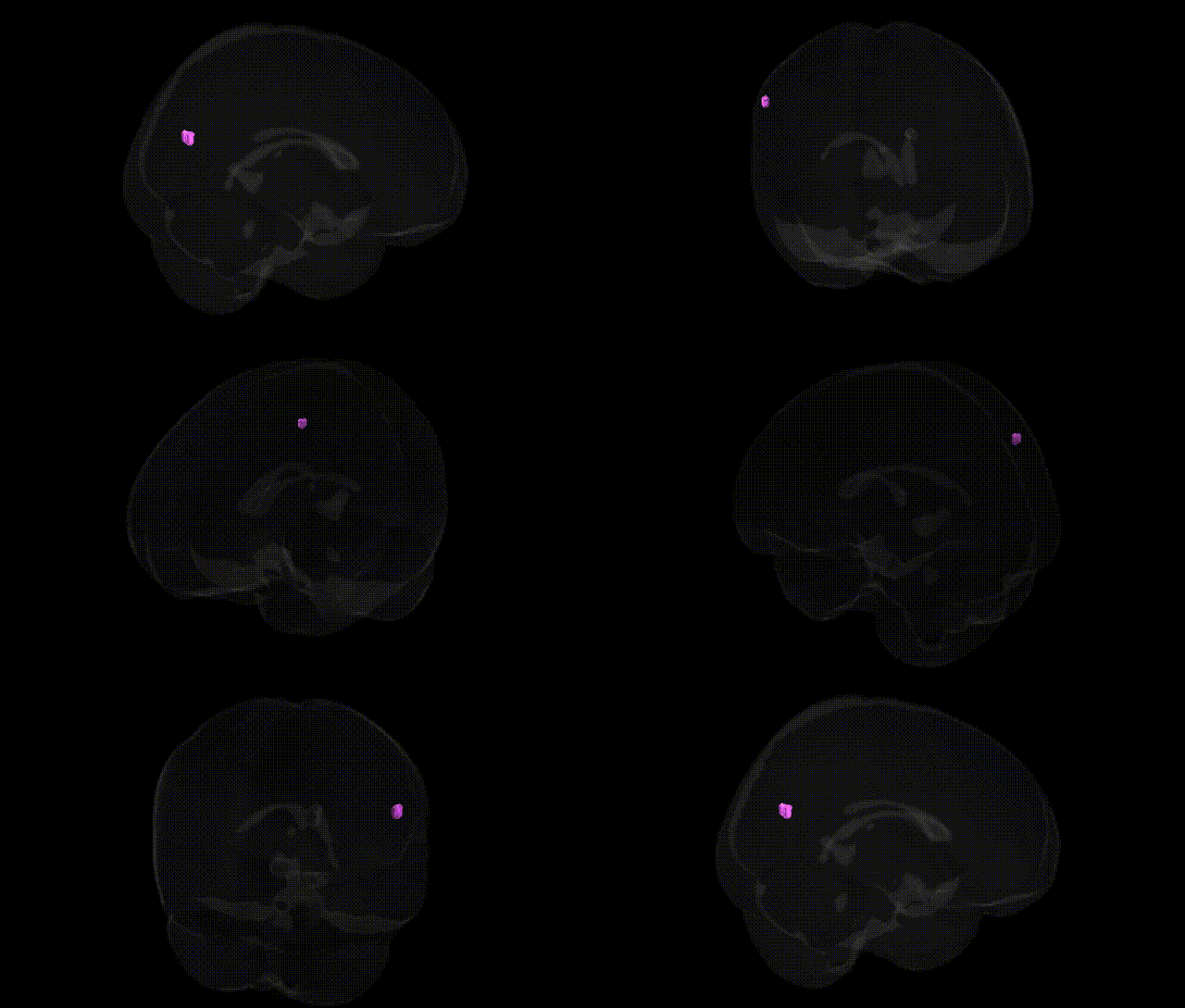
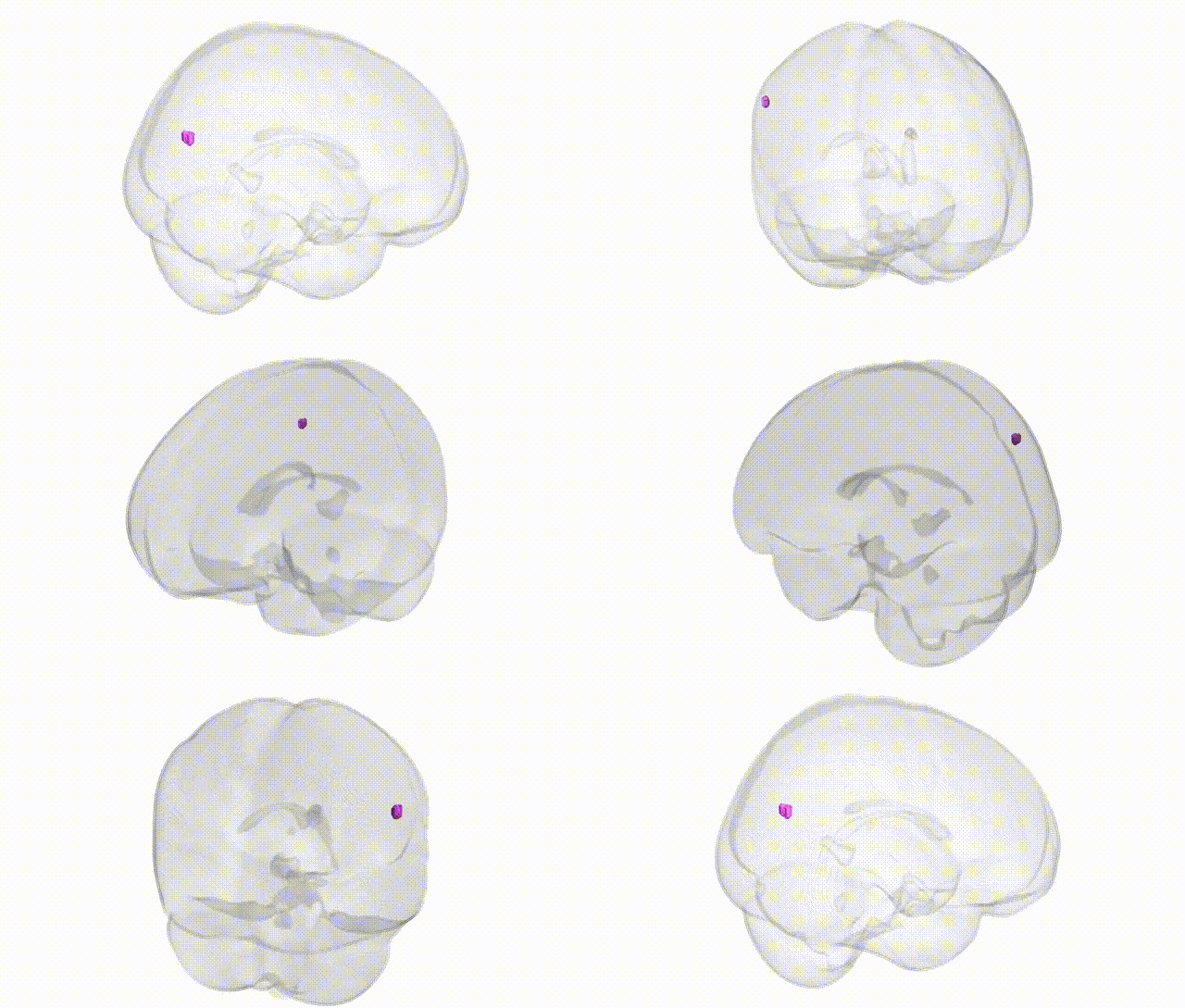
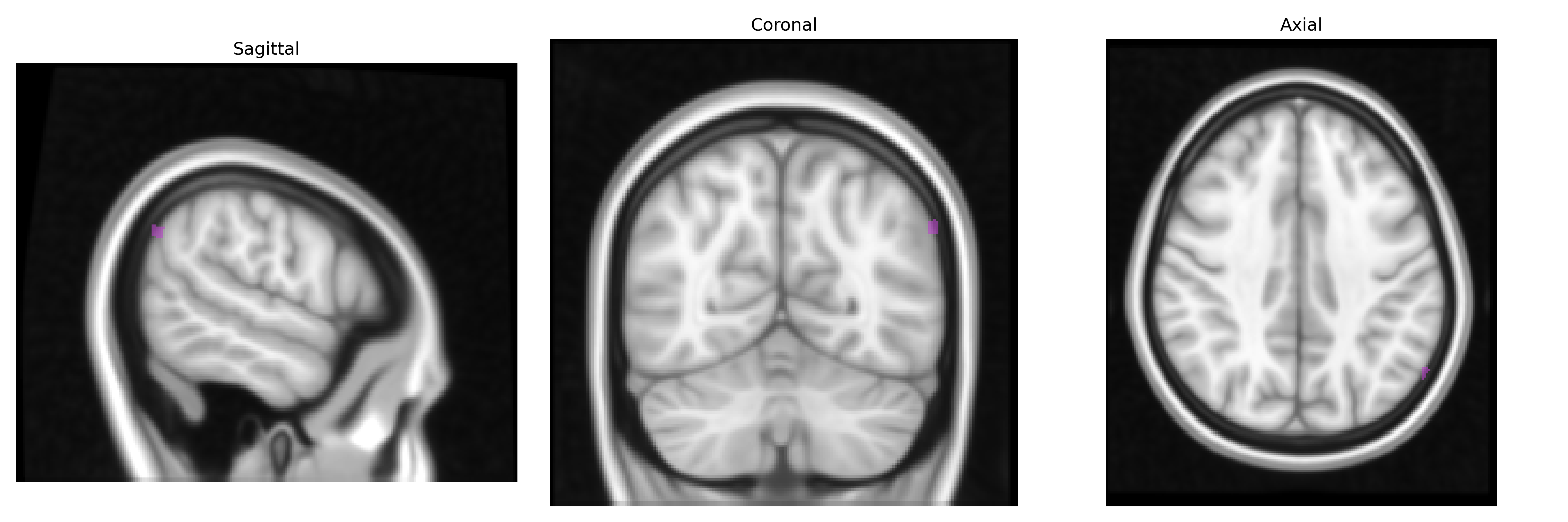
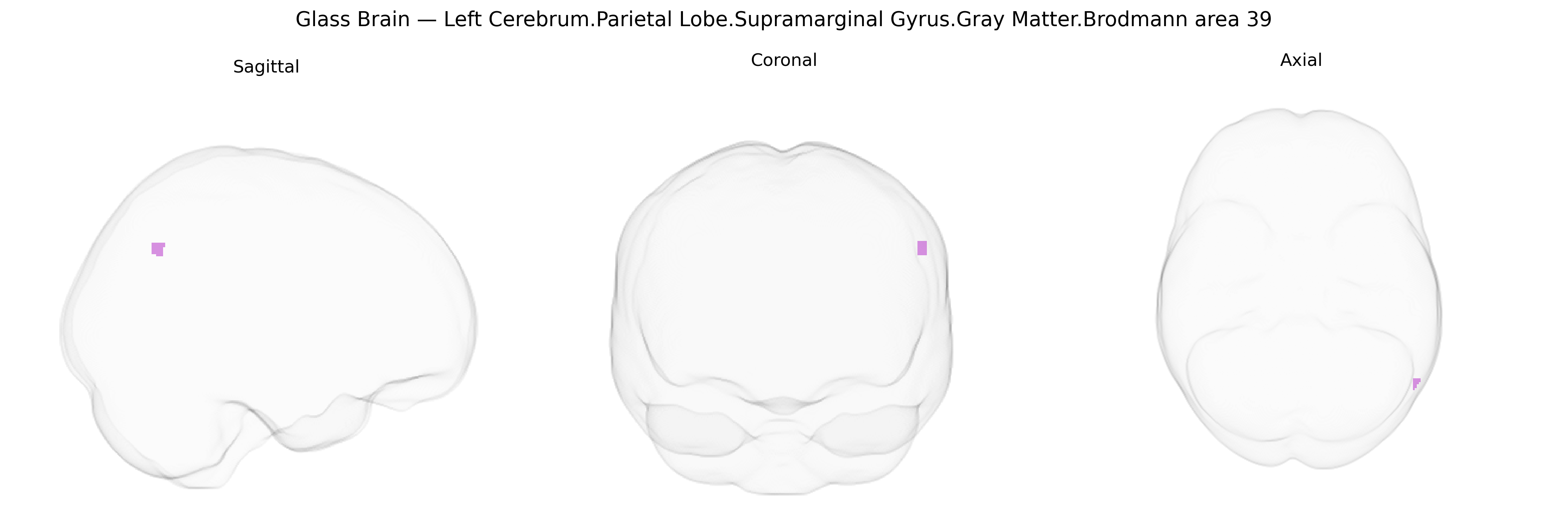
The bilateral Left Cerebrum.Parietal Lobe.Supramarginal Gyrus.Gray Matter.Brodmann area 39 corresponds primarily to the angular gyrus region in the inferior parietal lobule, a multimodal association cortex integrating visual, auditory, and somatosensory information. Cytoarchitectonically defined as Brodmann area 39, it participates in higher cognitive functions including language comprehension (particularly semantic processing), reading and writing, number processing, spatial cognition, theory of mind, and aspects of memory retrieval. This region forms part of the default mode and language networks, with strong connectivity to temporal, frontal, and occipital association areas. Lesions in this area are classically associated with Gerstmann syndrome (acalculia, agraphia, left-right disorientation, and finger agnosia) and can contribute to aphasia and other higher-order cognitive deficits. No direct link exists specifically for “Brodmann area 39” as a standalone supramarginal subdivision; a closely related and encompassing structure is the angular gyrus: Angular gyrus.
The supramarginal gyrus (SMG; Brodmann area 39) in the left parietal lobe, as defined in Talairach 1 mm atlases, has been implicated in multiple genetic and genome-wide association studies through its roles in language, reading, working memory, and social cognition. Structural and functional variation in this region has been associated with common variants in genes related to neuronal migration and synaptic plasticity (for example, CNTNAP2, FOXP2, KIAA0319, and DCDC2 in language and dyslexia studies), as well as polygenic scores for educational attainment and general cognitive ability, which often show effects on parietotemporal cortices including the SMG. GWAS of reading and spelling abilities, developmental dyslexia, and specific language impairment frequently report altered activation or gray matter volume in the left temporoparietal network encompassing the SMG, linking risk alleles in these genes to atypical structure or function in this area. Large-scale imaging–genetics consortia such as ENIGMA and UK Biobank have reported associations between common variants in genes involved in neurodevelopment, myelination, and synaptic signaling (e.g., variants near MIR137, MAPT, and other neuronal genes) and cortical thickness or surface area in inferior parietal regions that include the SMG. Additionally, the left SMG has appeared in imaging–genetic studies of schizophrenia, autism spectrum disorder, attention-deficit/hyperactivity disorder, and major depressive disorder, where risk alleles and polygenic risk scores correlate with altered gray matter or connectivity in this region, consistent with its role in higher-order cognition, language processing, and social-emotional integration.
Overview generated by GPT-4o (2026).
Region ID: 974
Hemisphere: bilateral
Atlas: Talairach labels 1mm
Full Quality Version: Download MP4
Full Quality Version: Download MP4
Wali Sidiqyar*, Gaurav Rudravaram*, Elyssa M. McMaster, Trent M. Schwartz, Adam M. Saunders, Kurt G. Schilling, Bennett A. Landman "Introducing SPINS: A Shared Public Visualization Library of Neuroanatomical Structures." Medical Imaging with Deep Learning- short paper
This resource is licensed under CC0 1.0 Universal (Public Domain).