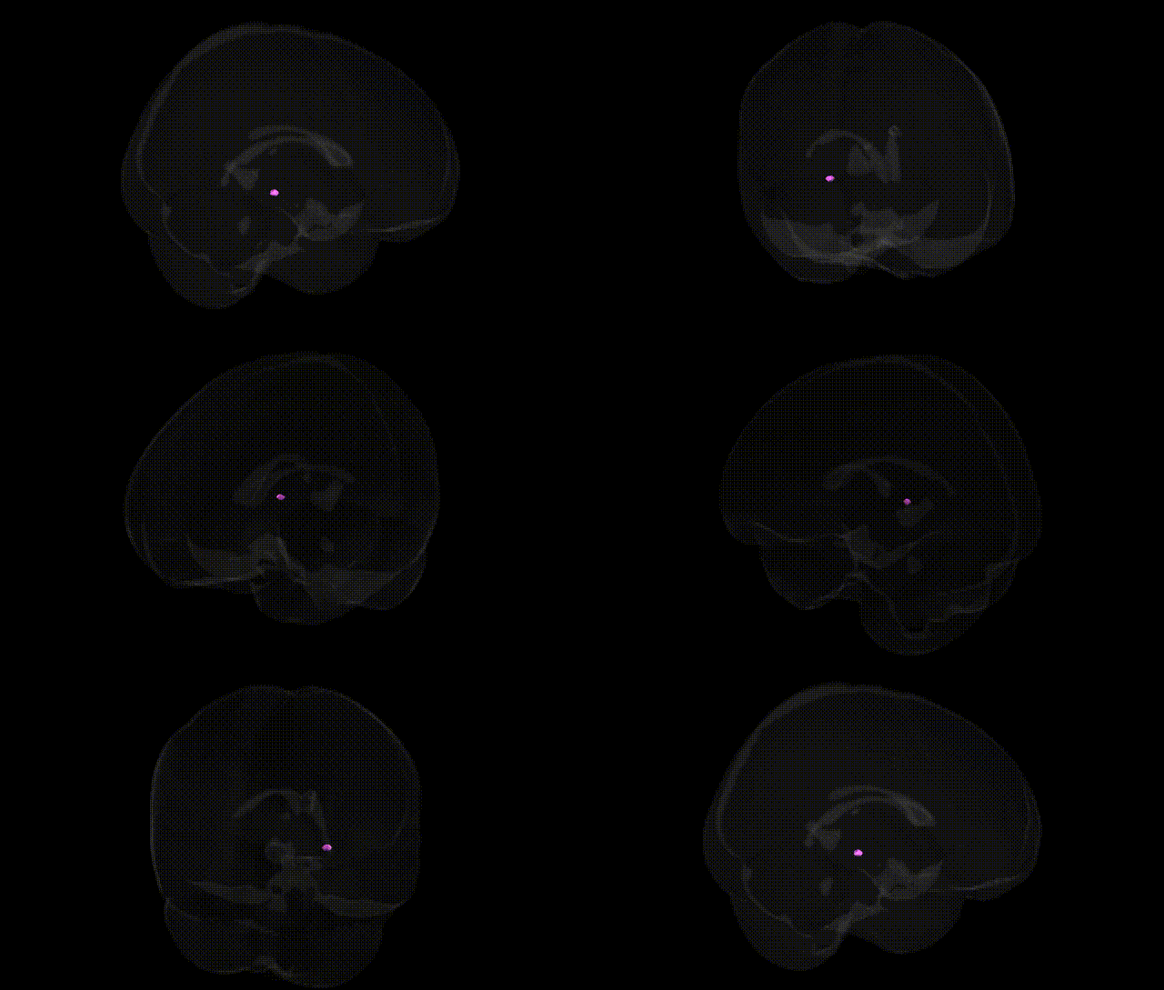
The bilateral Left Cerebrum.Sub-lobar.Gray Matter.Lateral Geniculum Body corresponds to the lateral geniculate nucleus (LGN) of the thalamus, a laminated relay nucleus within the subcortical gray matter that plays a central role in the primary visual pathway. It receives topographically organized retinal input via the optic tract and projects primarily to the primary visual cortex (V1, Brodmann area 17) through the optic radiations, preserving retinotopic organization and contributing to processing of contrast, spatial resolution, and color. The LGN contains magnocellular, parvocellular, and koniocellular layers, which support parallel processing of motion, form, and color information, and participates in modulatory loops with cortex and brainstem that influence visual attention and gain control. Functionally, it serves as a critical gate and integrator for visual signals, shaping early visual perception and contributing to conscious visual experience. Lateral geniculate nucleus
The lateral geniculate body (LGB), the thalamic relay of the visual pathway, shows genetic associations largely through imaging–genetics and GWAS of subcortical and thalamic volumes rather than region-specific disease GWAS; common variants in genes such as APOE, MAPT, and those within microtubule and synaptic pathways contribute to individual differences in thalamic and geniculate volumetry, and polygenic scores for neurodevelopmental and neurodegenerative traits partially explain variance in this structure. Large-scale ENIGMA and UK Biobank–based studies report that thalamic nuclei, including the lateral geniculate nucleus, share genetic influences with global brain morphometry and with psychiatric liability, particularly for schizophrenia, major depression, and bipolar disorder, consistent with thalamocortical dysconnectivity models. In neurodegenerative disease, genetic risk loci for Alzheimer’s disease and Parkinson’s disease (for example variants near APOE, BIN1, and SNCA) have been associated with altered thalamic and visual relay integrity on MRI and diffusion measures, implicating the LGB in the broader network of genetically mediated neurodegeneration. Visual pathway disorders such as glaucoma and multiple sclerosis involve geniculate atrophy and demyelination, and GWAS of these conditions—highlighting genes in immune regulation (HLA region, IL2RA, IL7R) and axonal integrity (e.g., neurofilament-related loci)—suggest that genetically driven vulnerability of myelinated visual pathways extends to the lateral geniculate body. Although few GWAS have been conducted with the LGB as a primary endpoint, convergent genetic evidence across subcortical volume, visual system disease, and psychiatric liability indicates that this structure lies at an intersection of polygenic influences on neurodevelopment, synaptic signaling, myelination, and neurodegeneration.
Overview generated by GPT-4o (2026).
Region ID: 497
Hemisphere: bilateral
Atlas: Talairach labels 1mm
Full Quality Version: Download MP4
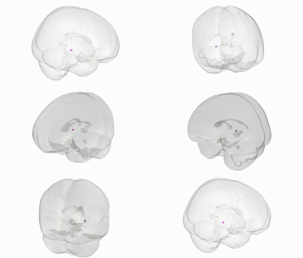
Full Quality Version: Download MP4
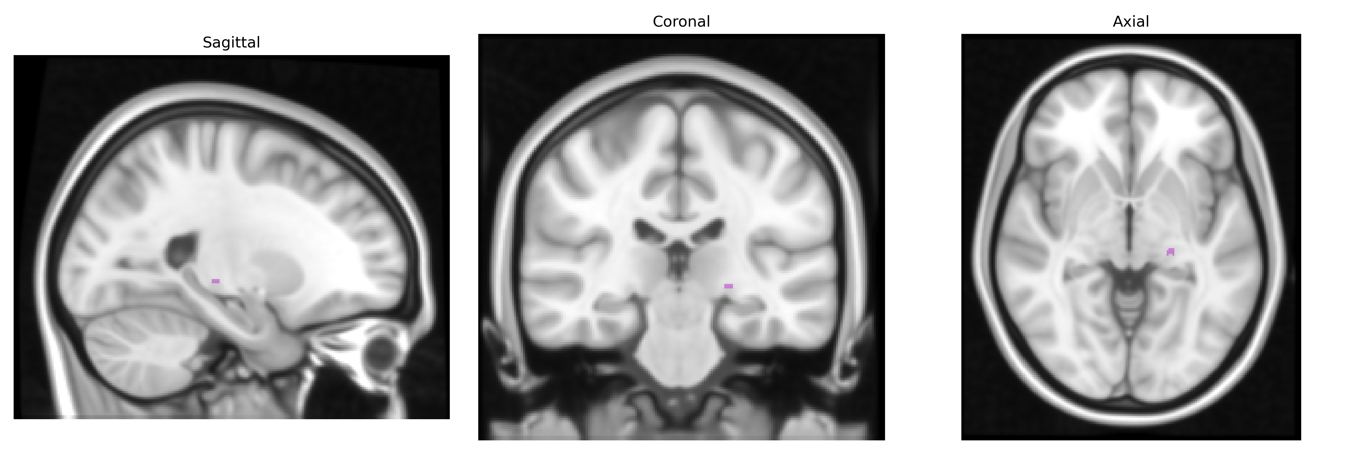
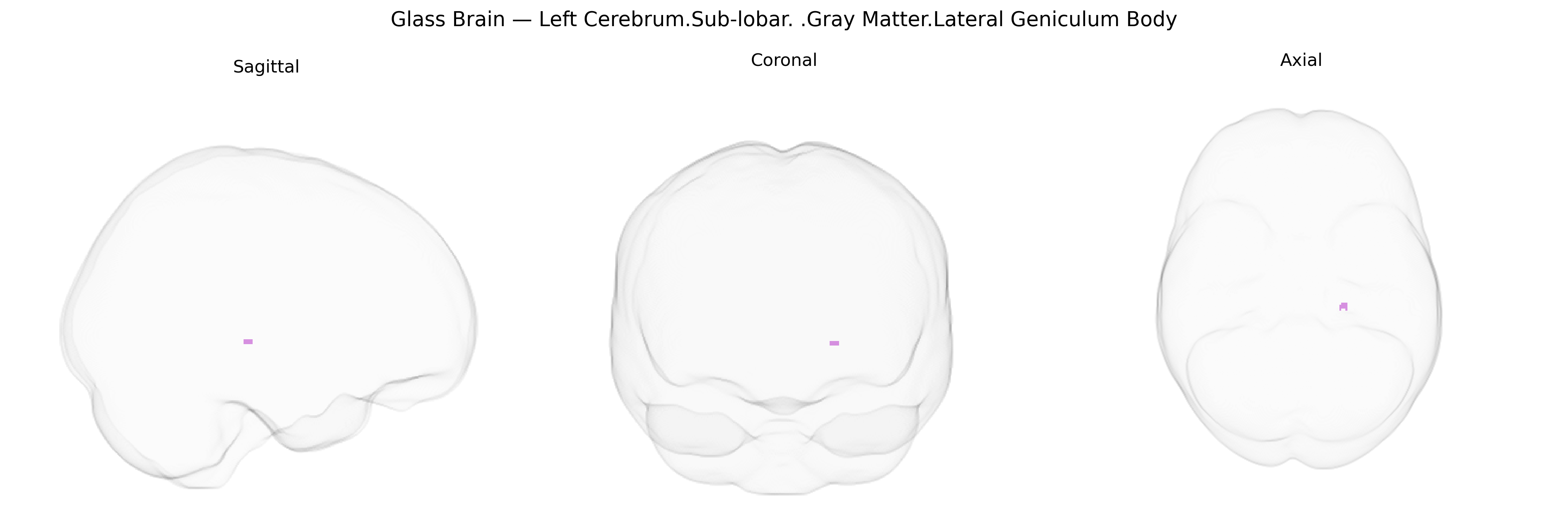
Wali Sidiqyar*, Gaurav Rudravaram*, Elyssa M. McMaster, Trent M. Schwartz, Adam M. Saunders, Kurt G. Schilling, Bennett A. Landman "Introducing SPINS: A Shared Public Visualization Library of Neuroanatomical Structures." Medical Imaging with Deep Learning- short paper
This resource is licensed under CC0 1.0 Universal (Public Domain).