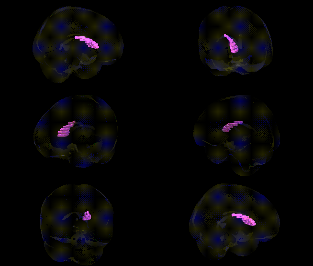
The bilateral Left Cerebrum.Sub-lobar.Caudate.Gray Matter.Caudate Body corresponds to the main, elongated middle portion of the caudate nucleus, a C-shaped gray matter structure in the subcortical (sub-lobar) region of the cerebral hemispheres that is a key component of the dorsal striatum and basal ganglia circuitry. The caudate body lies dorsal and lateral to the thalamus and medial to the internal capsule, and is composed predominantly of medium spiny GABAergic neurons that receive extensive excitatory glutamatergic inputs from frontal and associative cortices, as well as dopaminergic innervation from the substantia nigra pars compacta. Through its projections to the globus pallidus and substantia nigra, the caudate body participates in cortico-basal ganglia-thalamo-cortical loops that regulate motor planning, procedural and habit learning, action selection, and aspects of cognitive and emotional processing; dysfunction of this structure contributes to movement disorders such as Parkinson’s disease and Huntington’s disease, as well as certain neuropsychiatric conditions.
The bilateral caudate body gray matter in the left sub-lobar cerebrum has been implicated in multiple genetic and genome-wide association studies (GWAS) that link its volume, structure, and function to specific loci and polygenic architectures. Large-scale imaging-genetics consortia (e.g., ENIGMA, UK Biobank) have identified common variants near and within genes involved in neurodevelopment, synaptic signaling, and dopaminergic pathways—such as DRD2, PPP1R1B (DARPP-32), BDNF, and several loci in cell-adhesion and axon-guidance genes—as contributors to individual differences in caudate volume and shape. Polygenic risk for schizophrenia, bipolar disorder, and major depressive disorder correlates with altered caudate morphology, and GWAS of obsessive-compulsive disorder, Tourette syndrome, and attention-deficit/hyperactivity disorder implicate cortico-striatal circuits in which the caudate body is a key node. In movement disorders, Huntington’s disease is driven by a CAG repeat expansion in HTT that causes prominent caudate atrophy, while risk variants for Parkinson’s disease (e.g., in SNCA, LRRK2, GBA) affect striatal dopaminergic signaling and are associated with caudate functional and structural changes. Additionally, variants linked to general cognitive ability, educational attainment, and neuroticism show associations with caudate volume and connectivity, indicating that the genetic architecture of this region contributes to both neuropsychiatric risk and normal variation in cognition and personality.
Overview generated by GPT-4o (2026).
Region ID: 692
Hemisphere: bilateral
Atlas: Talairach labels 1mm
Full Quality Version: Download MP4
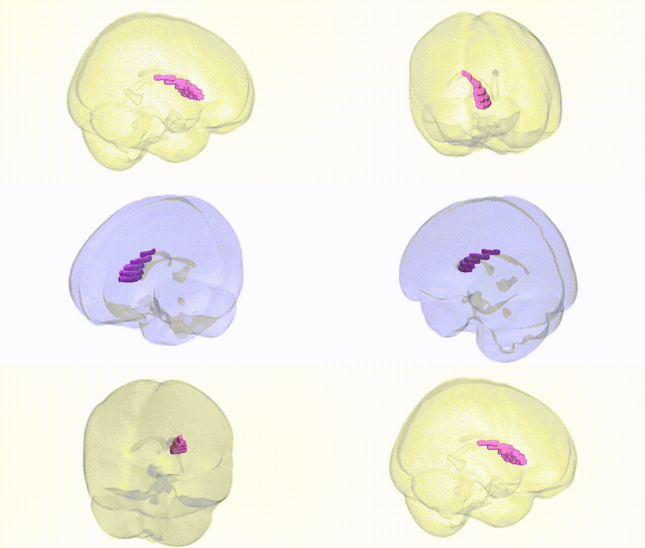
Full Quality Version: Download MP4
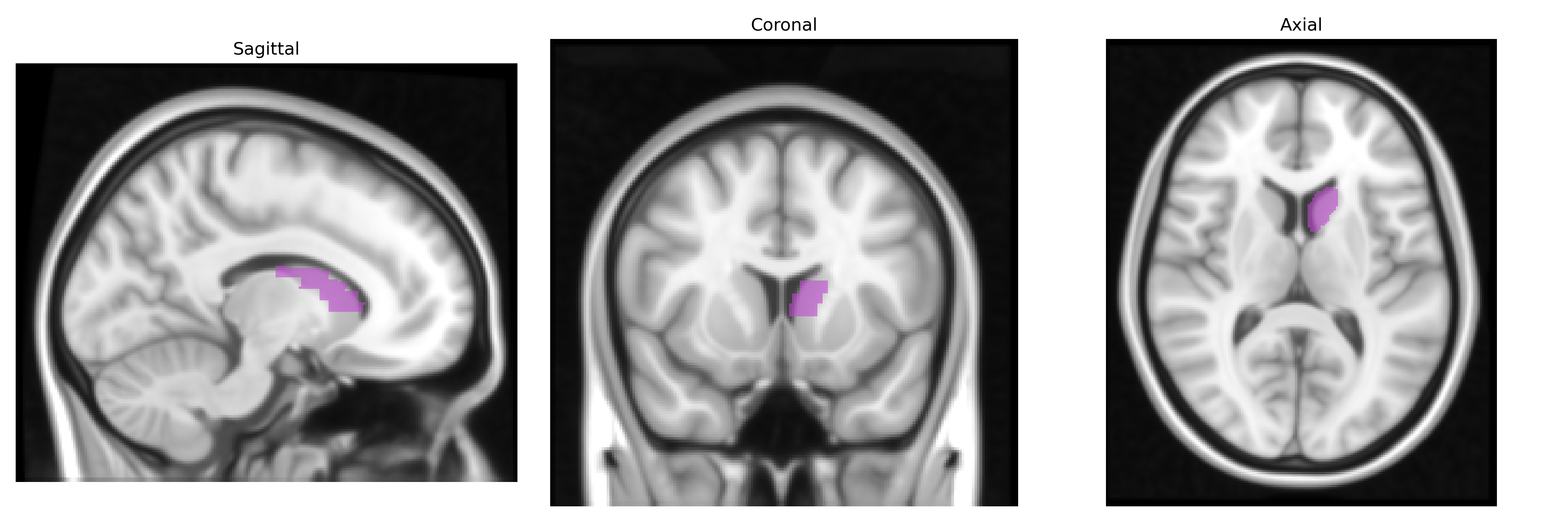
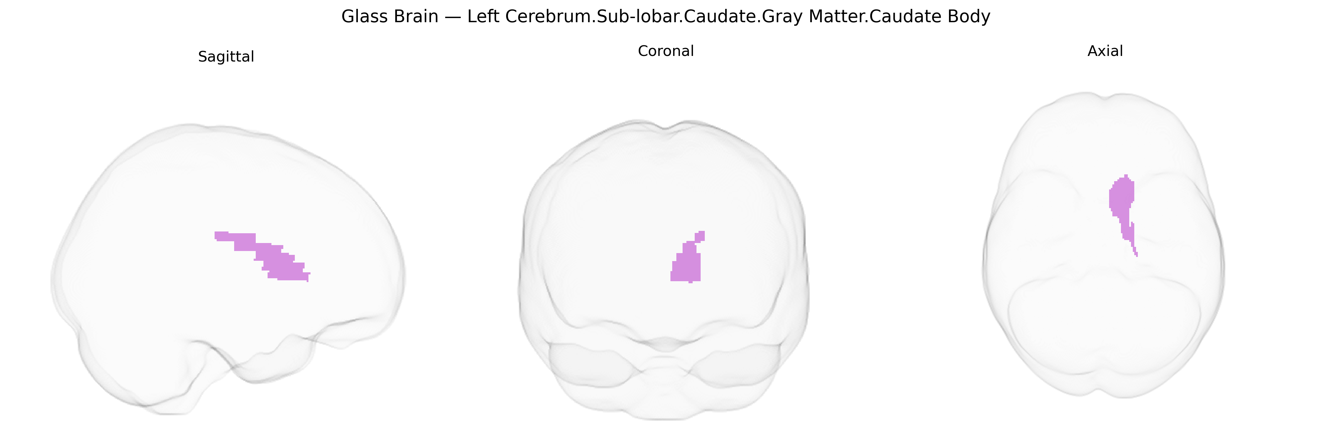
Wali Sidiqyar*, Gaurav Rudravaram*, Elyssa M. McMaster, Trent M. Schwartz, Adam M. Saunders, Kurt G. Schilling, Bennett A. Landman "Introducing SPINS: A Shared Public Visualization Library of Neuroanatomical Structures." Medical Imaging with Deep Learning- short paper
This resource is licensed under CC0 1.0 Universal (Public Domain).