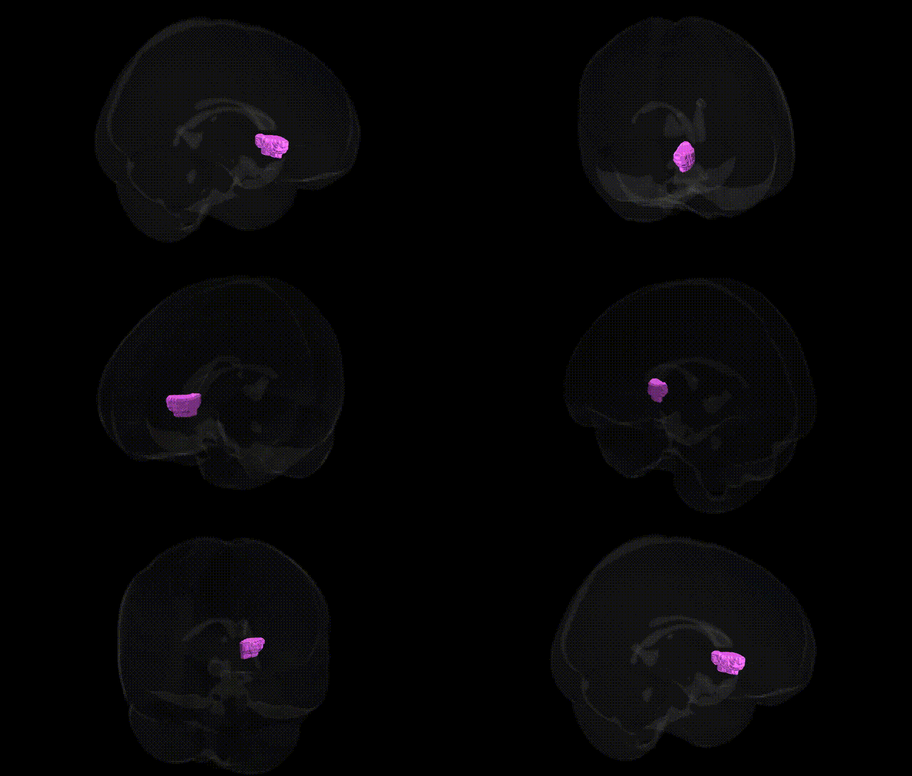
The bilateral Left Cerebrum.Sub-lobar.Caudate.Gray Matter.Caudate Head corresponds to the anterior portion of the caudate nucleus, a C-shaped gray matter structure located deep within the subcortical region of the cerebral hemispheres and forming part of the dorsal striatum within the basal ganglia. The caudate head lies adjacent to the lateral ventricle and is heavily interconnected with frontal cortical areas, particularly the prefrontal cortex, receiving dense glutamatergic inputs and projecting GABAergic outputs through basal ganglia circuits to the thalamus and back to cortex. It is critically involved in motor control, cognitive processes such as executive function and working memory, goal-directed behavior, and aspects of reward and learning. Pathologically, structural or functional alterations in the caudate head are implicated in movement disorders (e.g., Parkinson’s disease, Huntington’s disease), obsessive–compulsive disorder, and various neuropsychiatric conditions. Caudate nucleus
The bilateral caudate head (Talairach 1 mm, Left Cerebrum.Sub-lobar.Caudate.Gray Matter.Caudate Head) shows robust genetic influences on volume, shape, and connectivity, with SNP-heritability estimates often exceeding 0.7 in twin and family studies and numerous GWAS hits implicating neurodevelopmental and synaptic genes. Large-scale imaging-genetics consortia (e.g., ENIGMA, UK Biobank) have identified loci in and near genes such as C12orf75, FAT3, DAAM1, PTCH1, and SLC39A8, as well as polygenic contributions from pathways involved in axon guidance, neuronal migration, and dopaminergic signaling, that influence caudate volume and morphology. Genetically driven variation in caudate head structure and function has been associated with neuropsychiatric disorders and traits including schizophrenia, bipolar disorder, major depressive disorder, attention-deficit/hyperactivity disorder, autism spectrum disorder, obsessive–compulsive disorder, Tourette syndrome, and substance use, often via shared polygenic architectures linking case–control status with caudate volume or activation. Mendelian and risk variants impacting basal ganglia development (e.g., in HTT for Huntington’s disease, GNAL and other dystonia genes, and certain monogenic Parkinsonism genes) prominently affect the caudate, including its head, with early atrophy or dysfunction detectable by MRI. Additionally, genetic variants associated with cognitive performance, educational attainment, and personality traits (such as neuroticism and impulsivity) show correlations with caudate head volume or activation, suggesting that this region acts as a key, genetically modulated hub connecting cortico-striatal circuits underlying motivation, habit learning, reward processing, and executive control.
Overview generated by GPT-4o (2026).
Region ID: 464
Hemisphere: bilateral
Atlas: Talairach labels 1mm
Full Quality Version: Download MP4
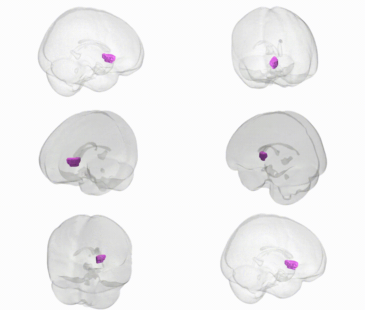
Full Quality Version: Download MP4
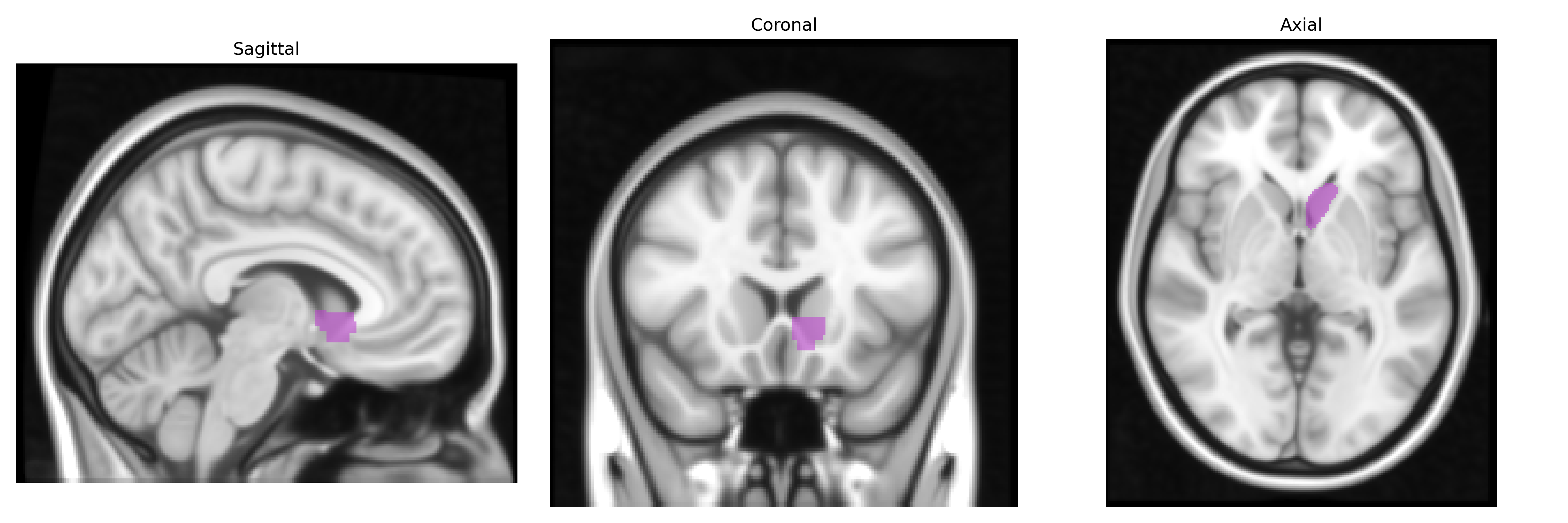
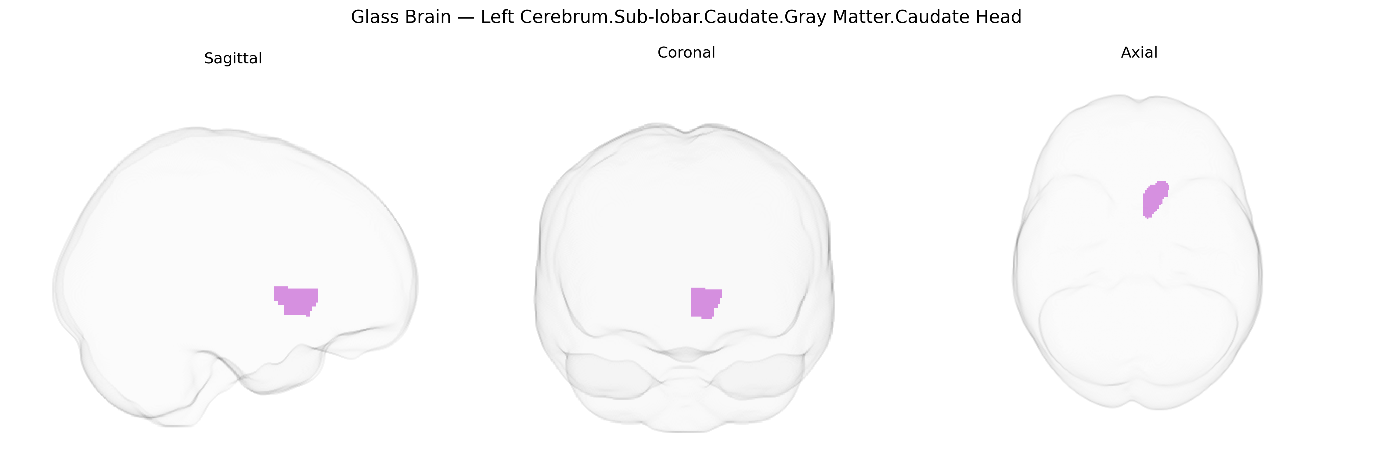
Wali Sidiqyar*, Gaurav Rudravaram*, Elyssa M. McMaster, Trent M. Schwartz, Adam M. Saunders, Kurt G. Schilling, Bennett A. Landman "Introducing SPINS: A Shared Public Visualization Library of Neuroanatomical Structures." Medical Imaging with Deep Learning- short paper
This resource is licensed under CC0 1.0 Universal (Public Domain).