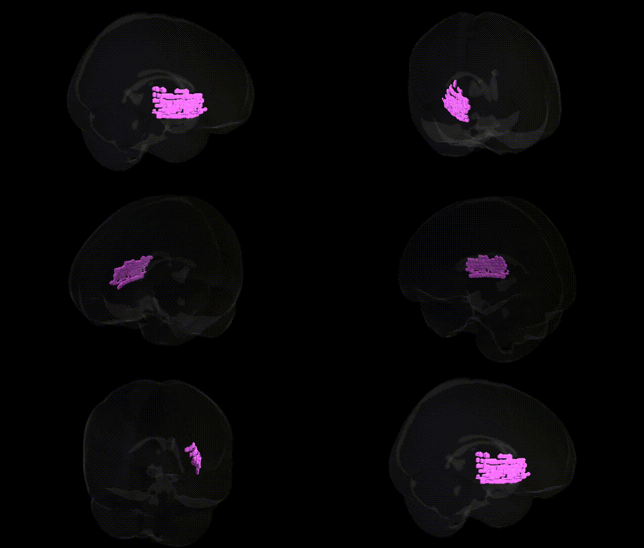
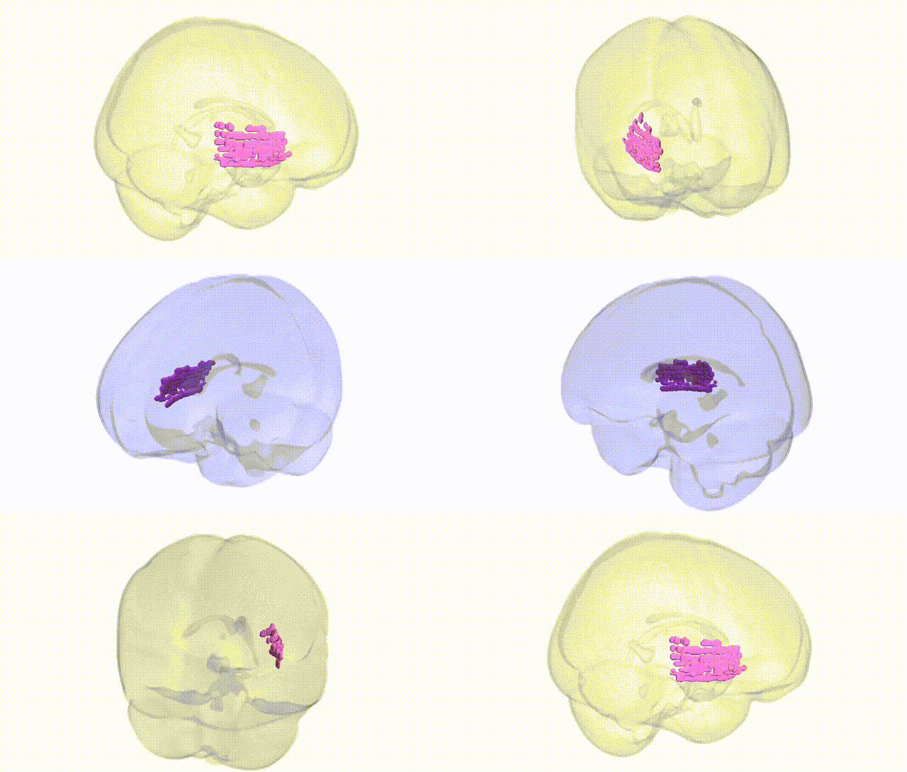
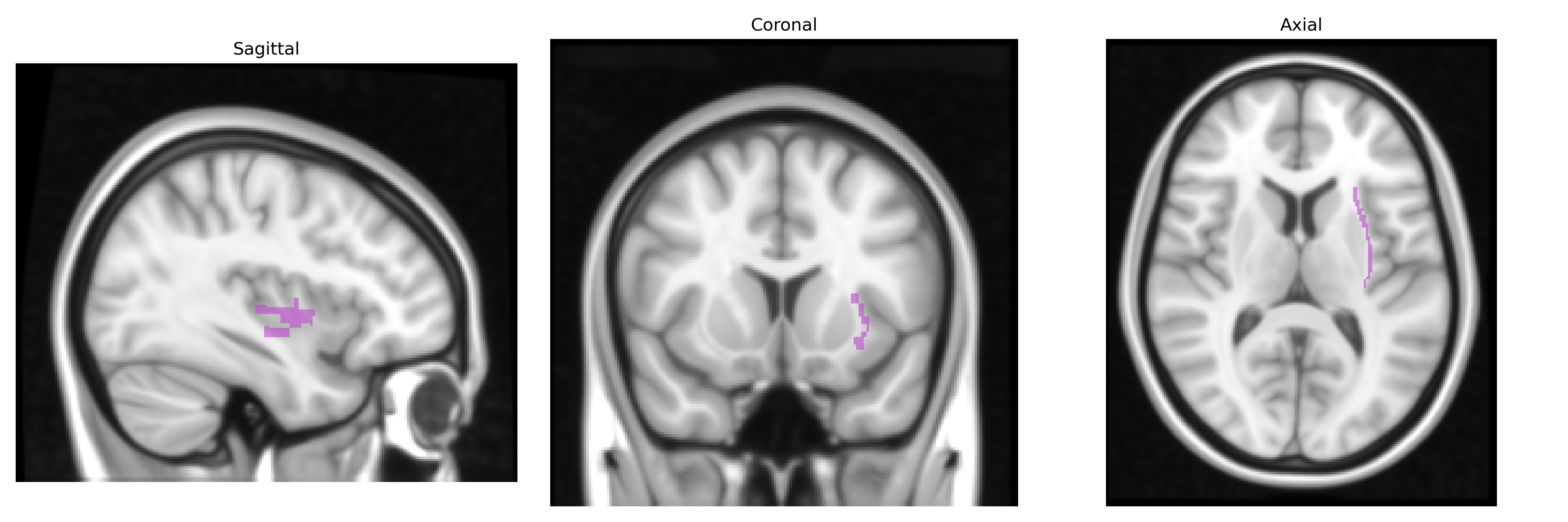
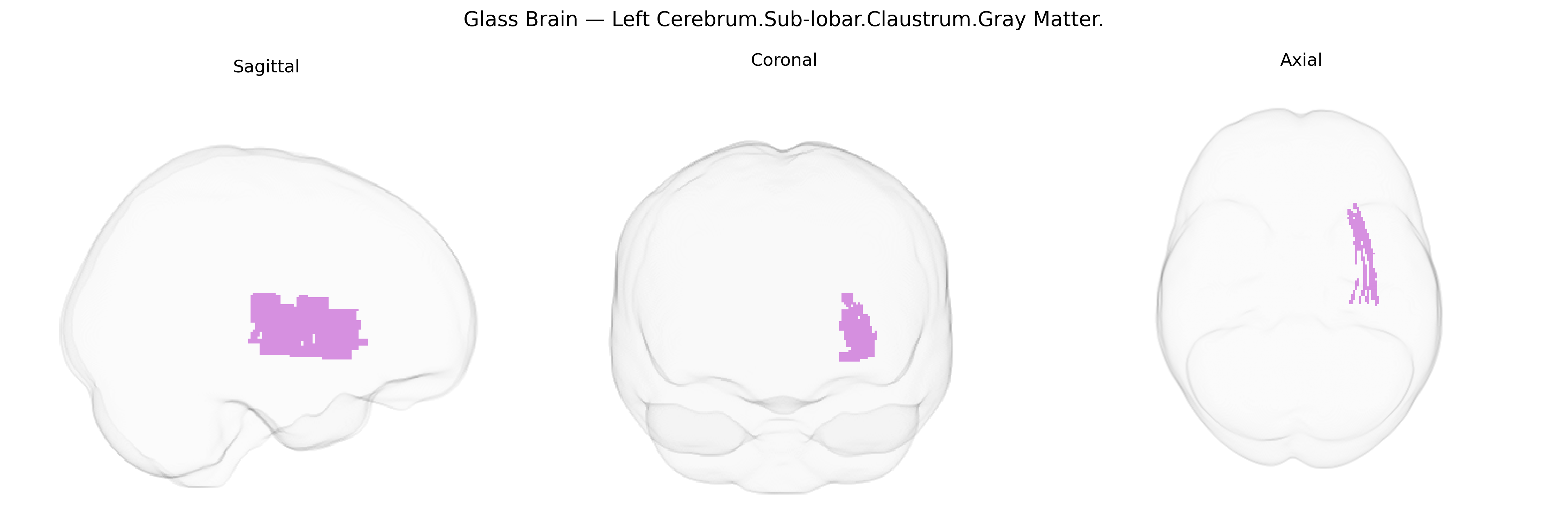
The bilateral Left Cerebrum.Sub-lobar.Claustrum.Gray Matter corresponds to the claustrum, a thin, irregular sheet of gray matter situated deep within the sub-lobar region of the cerebral hemisphere, interposed between the insular cortex medially and the putamen of the basal ganglia laterally. It is composed predominantly of densely packed neurons and is heavily interconnected with widespread cortical areas, including frontal, parietal, temporal, and occipital regions, suggesting a role in multimodal sensory integration, coordination of cortical activity, and possibly consciousness, attention, and salience processing. Although its precise function remains incompletely understood, the claustrum’s extensive reciprocal connections and strategic position imply a key role in synchronizing and modulating information flow across the cortex. Claustrum
The claustrum, a thin gray matter structure in the sub-lobar region of the left cerebrum, has limited direct GWAS evidence specifically targeting its gray-matter volume or thickness, but convergent genetic and imaging-genetic work implicates several pathways and disorders. Large-scale ENIGMA and UK Biobank imaging-GWAS studies that include subcortical and sub-lobar parcels suggest that gray-matter metrics in claustral or adjacent insulo-opercular regions show heritability and are influenced by polygenic variation in neurodevelopmental and synaptic genes, including loci related to neuronal migration, axon guidance, and cell-adhesion (for example genes in the neurexin/neuroligin and cadherin families, though often reported at broader regional or network levels rather than the claustrum specifically). The claustrum is frequently altered in structural MRI studies of schizophrenia, major depressive disorder, and bipolar disorder; twin and family-based designs indicate that volume reductions in this region partially reflect shared genetic liability with these psychiatric phenotypes, consistent with overlap with schizophrenia and depression polygenic risk scores. Mutations and risk variants in genes implicated in generalized epilepsy and temporal-lobe epilepsy (e.g., ion-channel and synaptic genes such as SCN-family and GABA-receptor genes) have been associated with seizure networks that prominently involve the claustrum, and case series report claustral lesions or dysfunction coinciding with genetic epilepsies, suggesting that genetic risk for epilepsy can manifest through claustral circuitry. In neurodegenerative disease, imaging-genetic analyses in Alzheimer’s and frontotemporal dementia cohorts show that risk alleles (e.g., APOE ε4 and variants near MAPT and GRN) are associated with atrophy patterns that extend into insulo-claustral regions, indicating that common dementia risk genes contribute to vulnerability of this structure. Overall, although few GWAS report claustrum-specific loci, the region appears genetically coupled to broader networks involved in psychiatric, epileptic, and neurodegenerative disorders through shared polygenic influences on cortical-subcortical connectivity, synaptic function, and neurodevelopment.
Overview generated by GPT-4o (2026).
Region ID: 449
Hemisphere: bilateral
Atlas: Talairach labels 1mm
Full Quality Version: Download MP4
Full Quality Version: Download MP4
Wali Sidiqyar*, Gaurav Rudravaram*, Elyssa M. McMaster, Trent M. Schwartz, Adam M. Saunders, Kurt G. Schilling, Bennett A. Landman "Introducing SPINS: A Shared Public Visualization Library of Neuroanatomical Structures." Medical Imaging with Deep Learning- short paper
This resource is licensed under CC0 1.0 Universal (Public Domain).