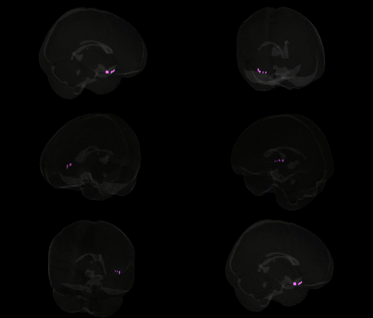
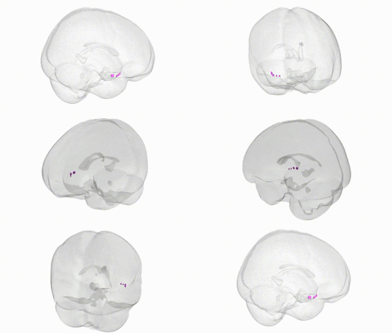
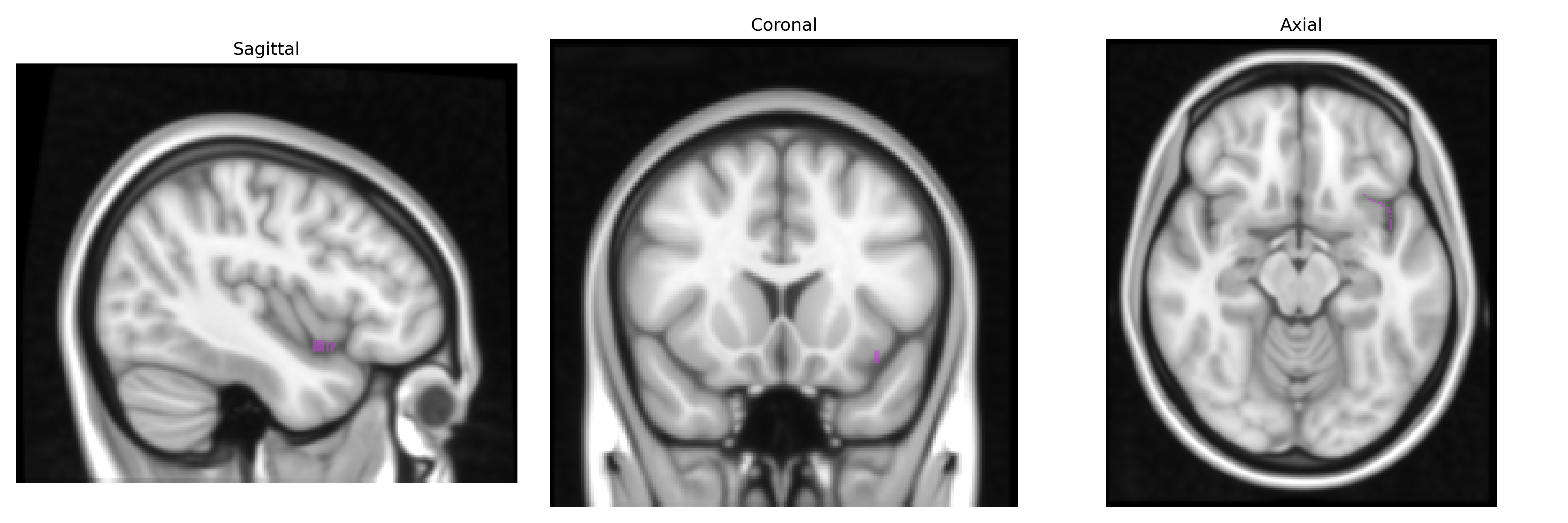
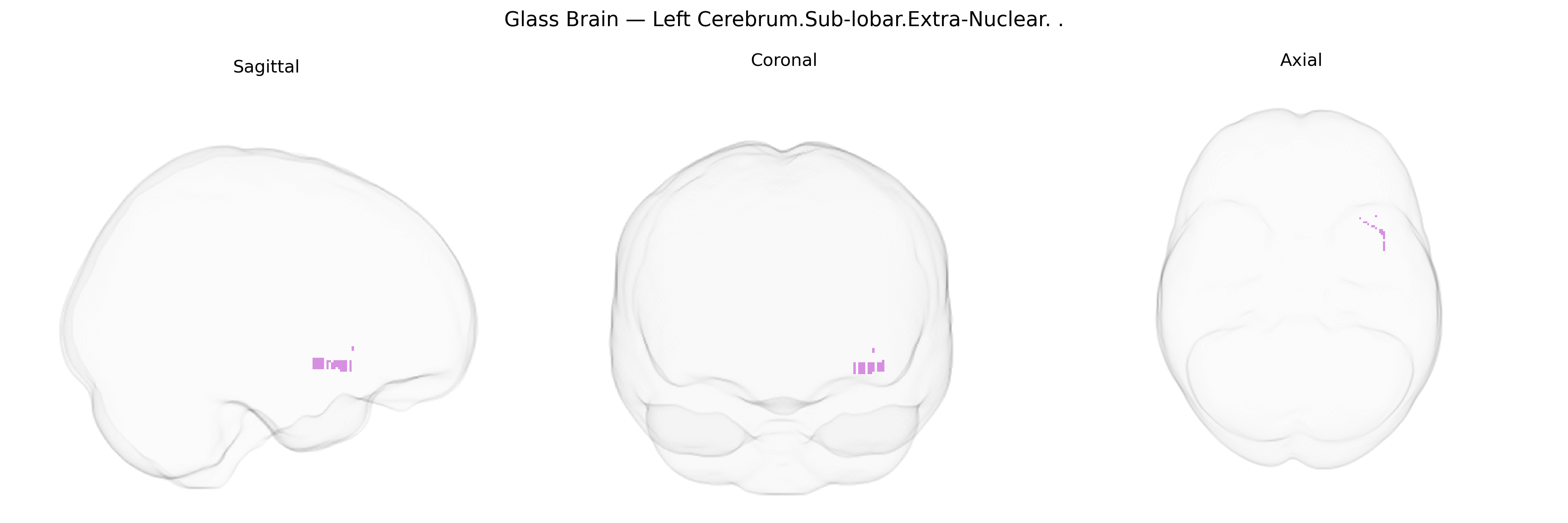
The bilateral Left Cerebrum.Sub-lobar.Extra-Nuclear region in the Talairach 1 mm atlas refers to subcortical, non-nuclear areas of the left cerebral hemisphere that lie outside the major gray-matter nuclei (such as the basal ganglia and thalamus) but within the sub-lobar compartment deep to the cerebral cortex. “Extra-nuclear” typically encompasses white matter tracts and transitional zones between cortical and nuclear structures, including portions of the internal capsule and surrounding deep white matter that integrate cortical signals with subcortical and brainstem nuclei. Functionally, these regions are crucial for long-range connectivity, supporting motor control, sensory integration, and higher cognitive processes by serving as conduits through which distributed neural networks communicate. There is no direct link for “Extra-Nuclear” as a named structure; a related and included structure is the Internal capsule.
The Talairach 1 mm “Left Cerebrum.Sub-lobar.Extra-Nuclear” label encompasses deep subcortical and periventricular white-matter regions (including areas traversed by projection and association fibers) that have been repeatedly implicated in imaging–genetics and GWAS studies of brain structure and neuropsychiatric disease. Large-scale GWAS of subcortical and white-matter volumes (e.g., ENIGMA and UK Biobank–based consortia) have identified common variants in genes involved in myelination and axonal integrity (such as MAG, PLP1, MBP, and other oligodendrocyte-related genes), neurodevelopmental patterning (e.g., variants near genes like NRG1, CNTNAP2, and NCAM1), and general brain growth and morphology (e.g., HMGA2, IGF1, and loci in 17q21.31) as contributors to individual differences in deep white-matter and sub-lobar volumes that intersect this “extra-nuclear” territory. Polygenic risk for schizophrenia, bipolar disorder, and major depression has been associated with reduced deep white-matter integrity and volume in this region and adjacent sub-lobar structures, while GWAS of stroke, leukoaraiosis, and white-matter hyperintensities highlight vascular and blood–brain barrier–related genes (e.g., COL4A1/2, HTRA1, and other small-vessel disease loci) in the same anatomical zone. Additional associations arise in GWAS of cognitive traits and educational attainment, where variants influencing global and regional white-matter microstructure (including tracts coursing through extra-nuclear sub-lobar zones) partially mediate genetic effects on processing speed and general cognitive ability. Overall, the genetic architecture of the Left Cerebrum.Sub-lobar.Extra-Nuclear region reflects a convergence of neurodevelopmental, myelination, vascular, and psychiatric risk pathways, although precise gene–region mappings are typically inferred from broader subcortical and white-matter parcels rather than this atlas label alone.
Overview generated by GPT-4o (2026).
Region ID: 378
Hemisphere: bilateral
Atlas: Talairach labels 1mm
Full Quality Version: Download MP4
Full Quality Version: Download MP4
Wali Sidiqyar*, Gaurav Rudravaram*, Elyssa M. McMaster, Trent M. Schwartz, Adam M. Saunders, Kurt G. Schilling, Bennett A. Landman "Introducing SPINS: A Shared Public Visualization Library of Neuroanatomical Structures." Medical Imaging with Deep Learning- short paper
This resource is licensed under CC0 1.0 Universal (Public Domain).