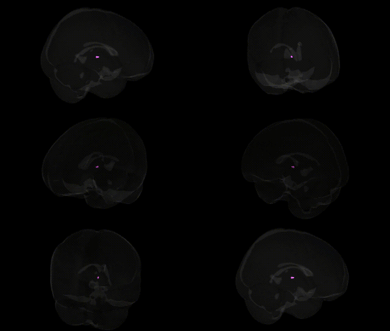
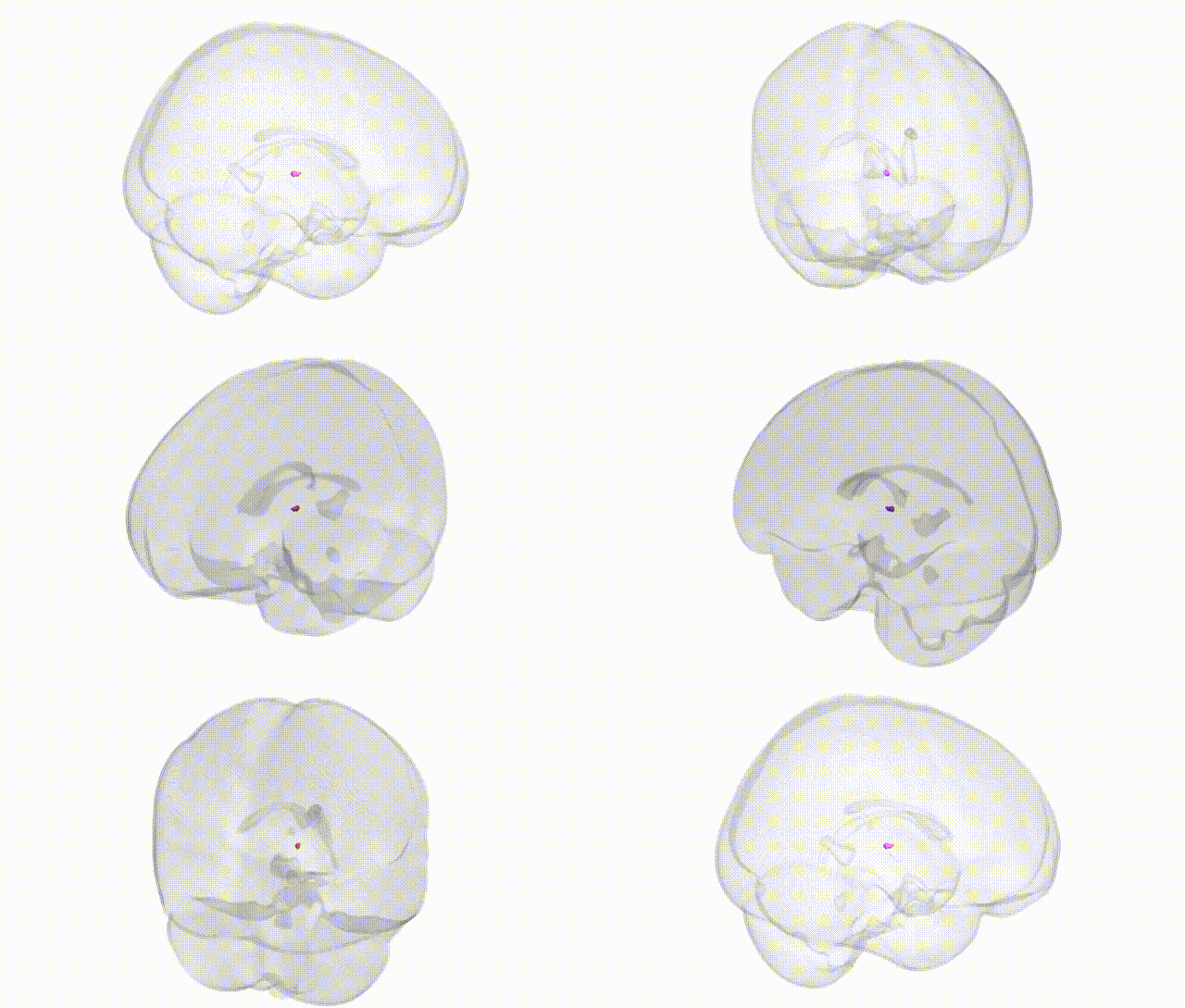
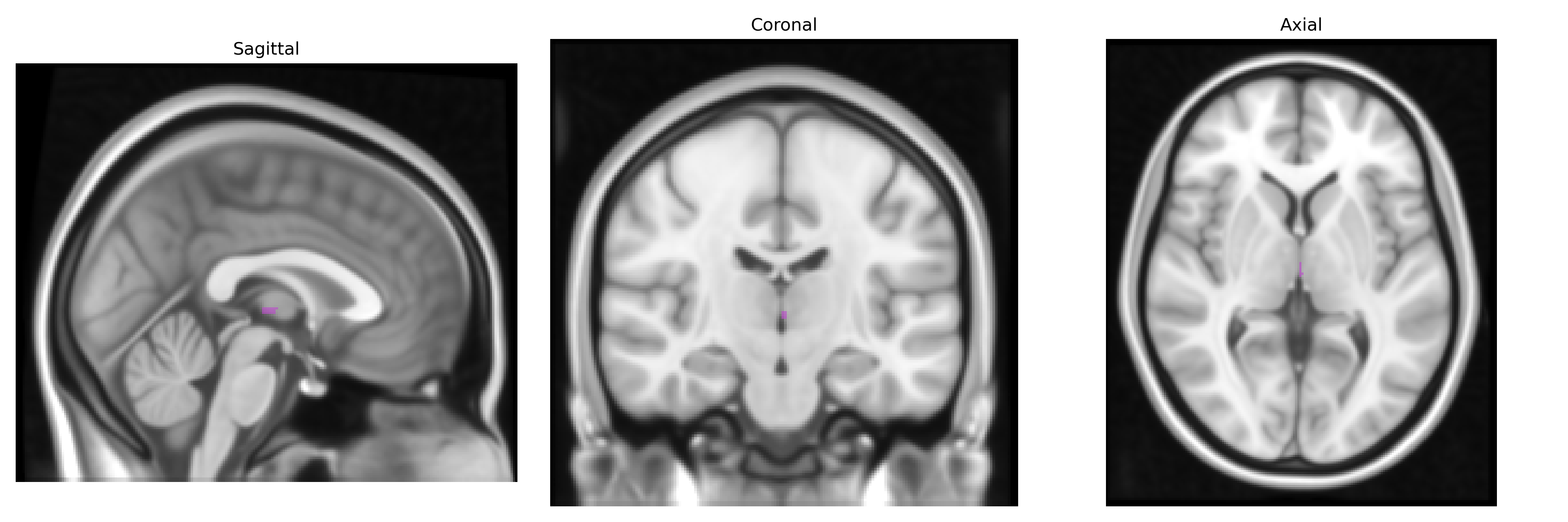
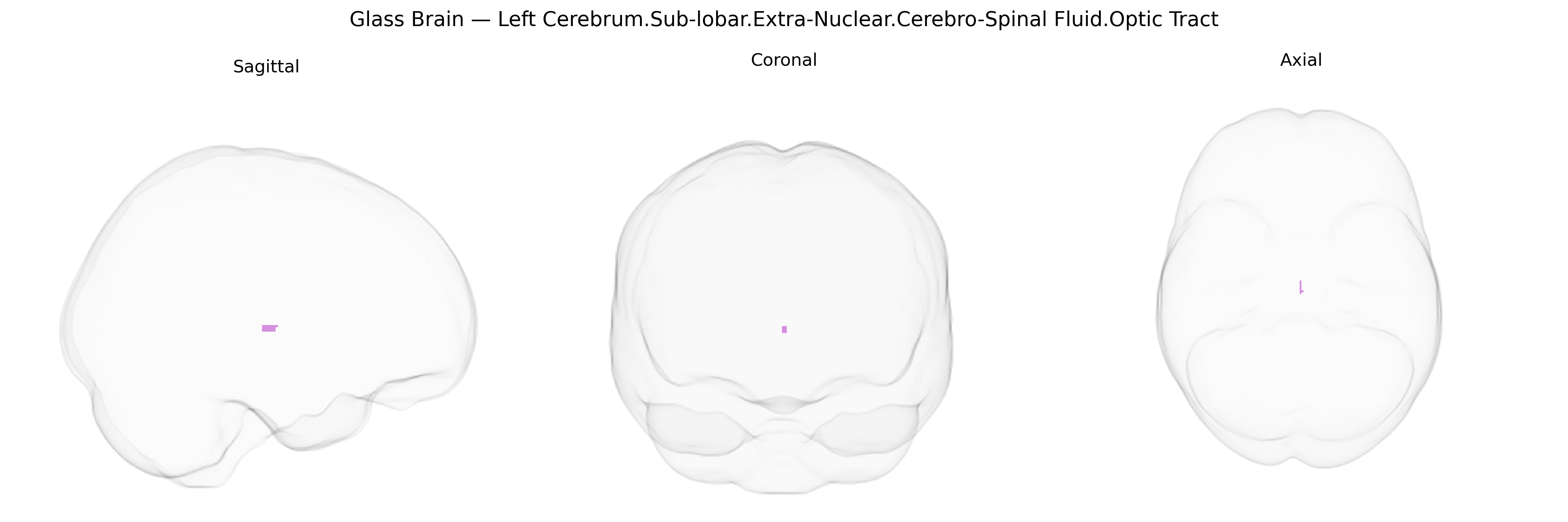
The bilateral Left Cerebrum.Sub-lobar.Extra-Nuclear.Cerebro-Spinal Fluid.Optic Tract label in the Talairach 1 mm atlas refers to cerebrospinal fluid (CSF)-filled spaces localized adjacent to the optic tract within the sublobar (deep) region of the cerebrum, outside the major nuclear (grey-matter) complexes. In this context, “extra-nuclear” indicates CSF rather than parenchymal tissue, and the region typically corresponds to fluid-containing compartments such as the subarachnoid space or ventricular recesses that course near the optic pathway as it extends from the optic chiasm toward the lateral geniculate nucleus. These CSF spaces contribute to mechanical protection, metabolic support, and clearance functions around the deep visual pathways and are intimately related to, but distinct from, the myelinated fiber bundle of the Optic tract.
The bilateral left sub-lobar extra-nuclear cerebrospinal fluid region around the optic tract, as defined in the Talairach 1 mm atlas, overlaps anatomically with white-matter pathways of the optic tract and adjacent periventricular/CSF spaces, but genetic association studies rarely target this specific label; instead, GWAS of brain imaging traits have identified variants influencing optic tract size, white-matter microstructure (e.g., fractional anisotropy), and periventricular CSF volumes as part of broader visual pathway and global brain morphology phenotypes. Large neuroimaging GWAS consortia such as ENIGMA and UK Biobank have reported heritability and polygenic influences on optic radiation and related tracts, implicating genes involved in axonal guidance, myelination, and neurodevelopment (for example, loci near genes like NRG1, MAG, and those in oligodendrocyte-linked pathways), though the associations are usually reported for composite white-matter or visual pathway measures rather than for the Talairach “Extra-Nuclear CSF/Optic Tract” label itself. Clinically, Mendelian disorders and complex traits affecting the optic nerve and tract—such as Leber hereditary optic neuropathy (MT-ND gene mutations), dominant optic atrophy (OPA1), and multiple sclerosis susceptibility loci identified by GWAS—can lead to structural and microstructural alterations of the optic tract region detectable on MRI, but these conditions are not mapped to a single atlas-defined CSF label. Overall, current evidence supports substantial genetic contributions to the structure and integrity of the optic tract and surrounding white matter, yet no robust, widely replicated GWAS has focused explicitly on the Talairach bilateral left sub-lobar extra-nuclear CSF–optic tract region as a distinct quantitative trait, and most findings derive from larger-scale visual pathway or global white-matter analyses.
Overview generated by GPT-4o (2026).
Region ID: 638
Hemisphere: bilateral
Atlas: Talairach labels 1mm
Full Quality Version: Download MP4
Full Quality Version: Download MP4
Wali Sidiqyar*, Gaurav Rudravaram*, Elyssa M. McMaster, Trent M. Schwartz, Adam M. Saunders, Kurt G. Schilling, Bennett A. Landman "Introducing SPINS: A Shared Public Visualization Library of Neuroanatomical Structures." Medical Imaging with Deep Learning- short paper
This resource is licensed under CC0 1.0 Universal (Public Domain).