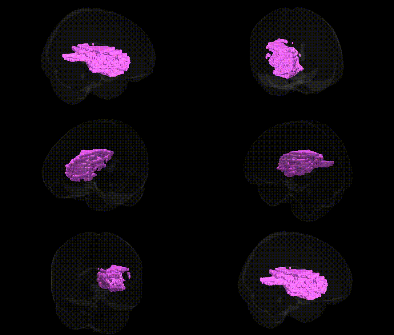
The bilateral Left Cerebrum.Sub-lobar.Extra-Nuclear.White Matter, as defined in the Talairach 1 mm atlas, encompasses deep subcortical white matter tracts situated beneath the cortical mantle and outside the major nuclear (gray matter) complexes such as the basal ganglia and thalamus. This region contains long association, projection, and commissural fibers that interconnect cortical areas with one another and with subcortical and brainstem structures, supporting efficient communication required for sensorimotor integration, higher cognition, and language processing. It includes portions of major pathways such as the internal capsule and surrounding deep white matter fields, serving as a critical substrate for rapid signal conduction and functional integration across distributed neural networks. There is no direct link for this exact atlas label; see the related structure Cerebral white matter.
The bilateral Left Cerebrum.Sub-lobar.Extra-Nuclear.White Matter region, as defined in the Talairach 1 mm atlas, corresponds largely to deep fronto-subcortical and periventricular white matter tracts rather than a single functionally discrete structure, and current genetic literature rarely targets it by this exact label; instead, genome-wide association studies (GWAS) typically examine broader white matter measures such as fractional anisotropy (FA), mean diffusivity (MD), or specific tracts (e.g., anterior limb of internal capsule, superior longitudinal fasciculus), which anatomically traverse or neighbor this extra-nuclear white matter. Large imaging-genetics consortia (e.g., ENIGMA, UK Biobank) have identified robust associations between variants in genes involved in axonal guidance and myelination (such as NTRK1/2, CNTN4, MAG, MBP, and genes within the 17q21.31 region including MAPT) and white matter microstructure in fronto-subcortical regions, as well as polygenic overlap between global and regional white matter integrity and neuropsychiatric disorders including schizophrenia, bipolar disorder, major depressive disorder, ADHD, and autism spectrum disorder. Moreover, loci in vascular and inflammatory genes (e.g., COL4A1/2, HTRA1, NOTCH3, genes in the 9p21 region) have been associated with deep and periventricular white matter hyperintensities, lacunar infarcts, and cerebral small vessel disease, which preferentially affect sub-lobar white matter and are genetically correlated with stroke, hypertension, and cognitive decline. While no major study has reported a locus uniquely tied to the Talairach-defined “extra-nuclear” white matter label specifically, convergent imaging-GWAS evidence indicates that genetic variation influencing myelination, axonal structure, vascular integrity, and neurodevelopment contributes to interindividual differences in the microstructure and lesion burden of these deep white matter pathways, in turn modulating risk for cognitive performance, dementia, mood and psychotic disorders, and cerebrovascular outcomes.
Overview generated by GPT-4o (2026).
Region ID: 347
Hemisphere: bilateral
Atlas: Talairach labels 1mm
Full Quality Version: Download MP4
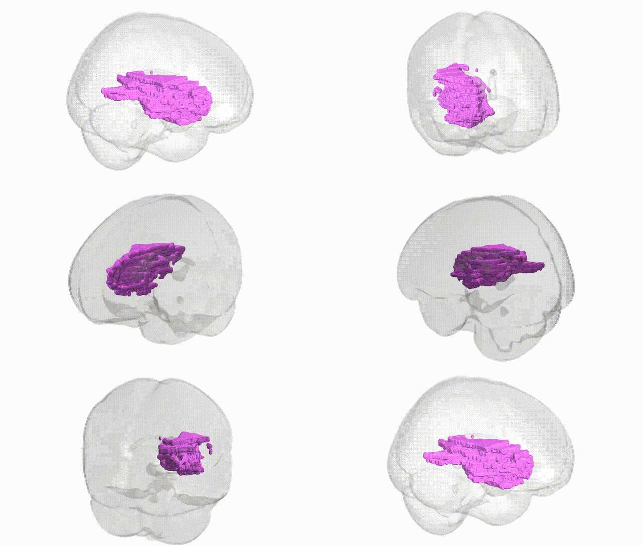
Full Quality Version: Download MP4
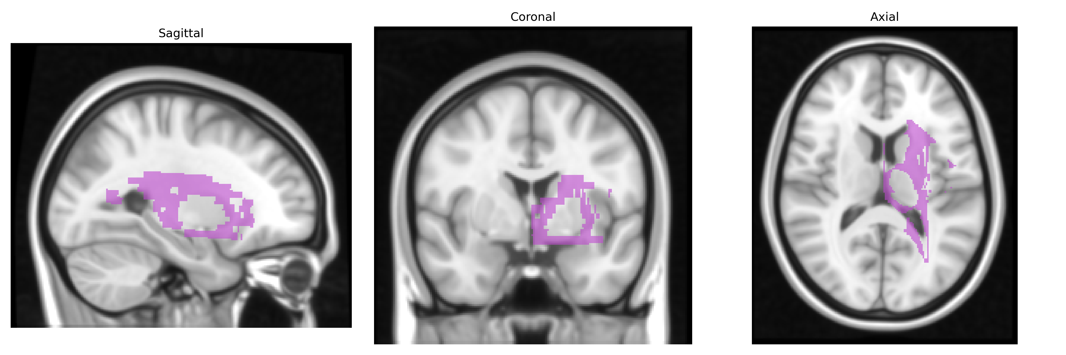
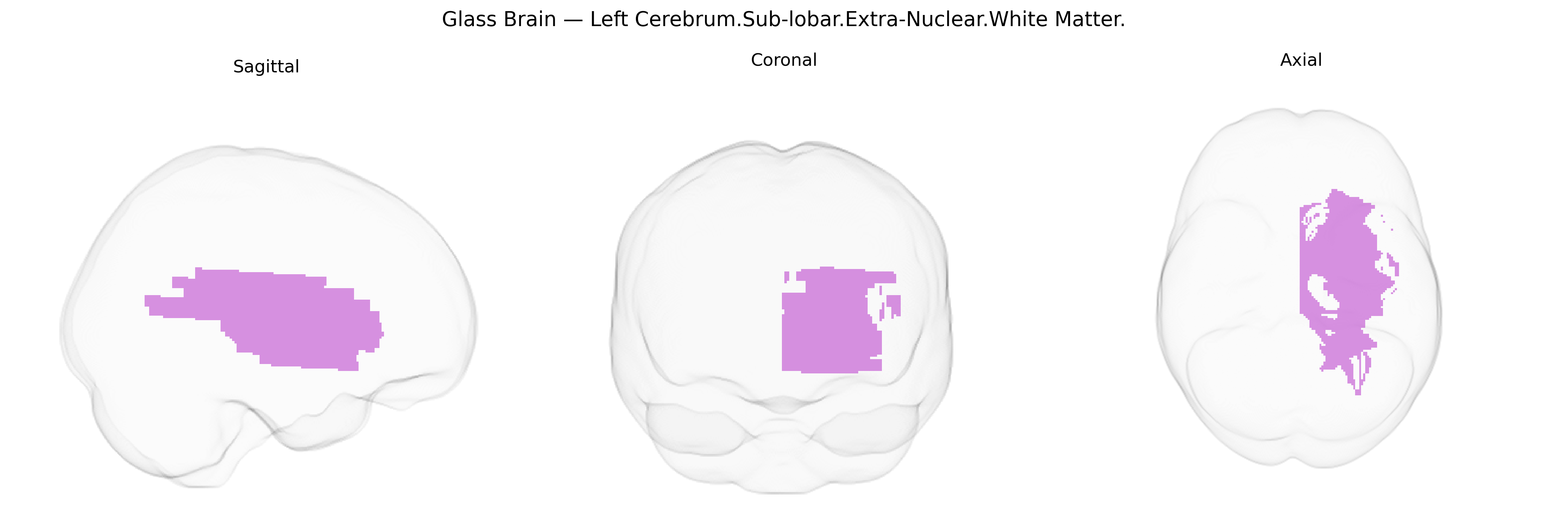
Wali Sidiqyar*, Gaurav Rudravaram*, Elyssa M. McMaster, Trent M. Schwartz, Adam M. Saunders, Kurt G. Schilling, Bennett A. Landman "Introducing SPINS: A Shared Public Visualization Library of Neuroanatomical Structures." Medical Imaging with Deep Learning- short paper
This resource is licensed under CC0 1.0 Universal (Public Domain).