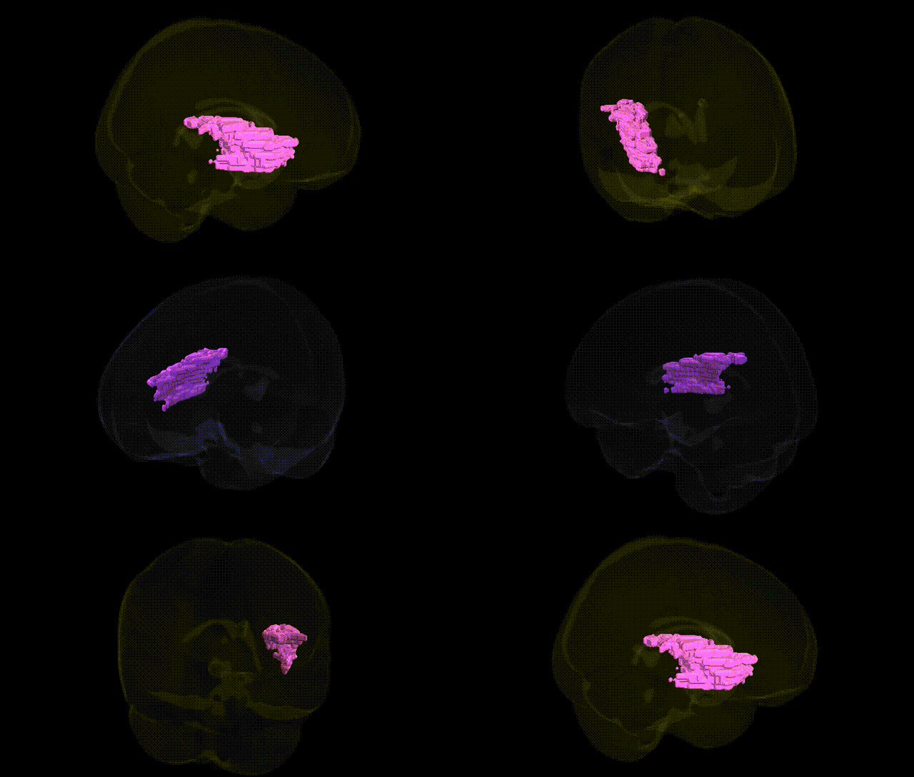
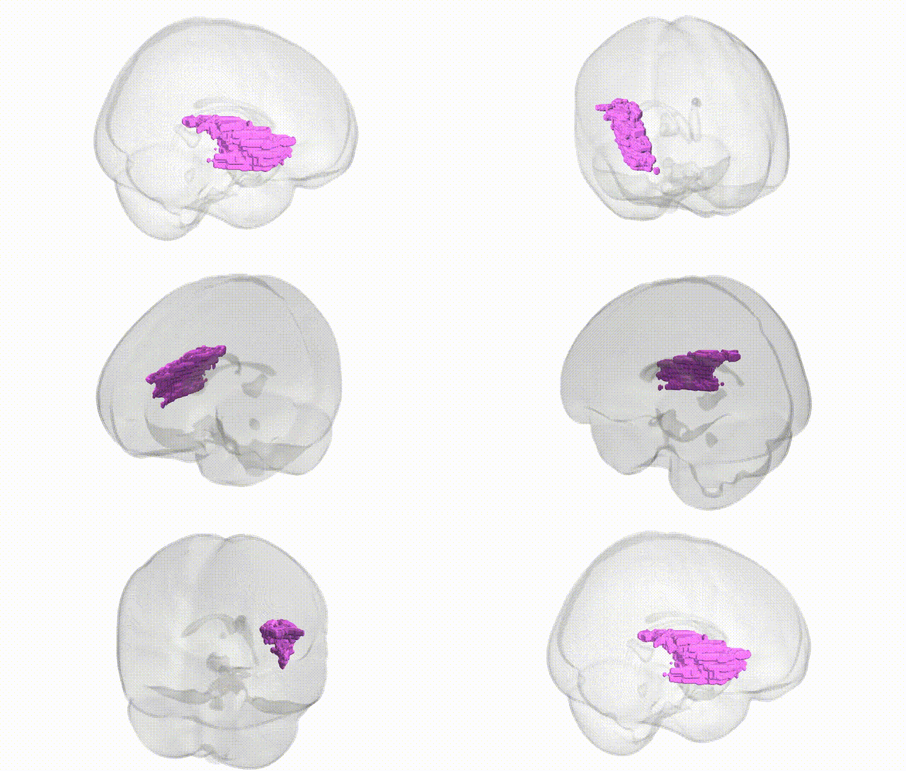
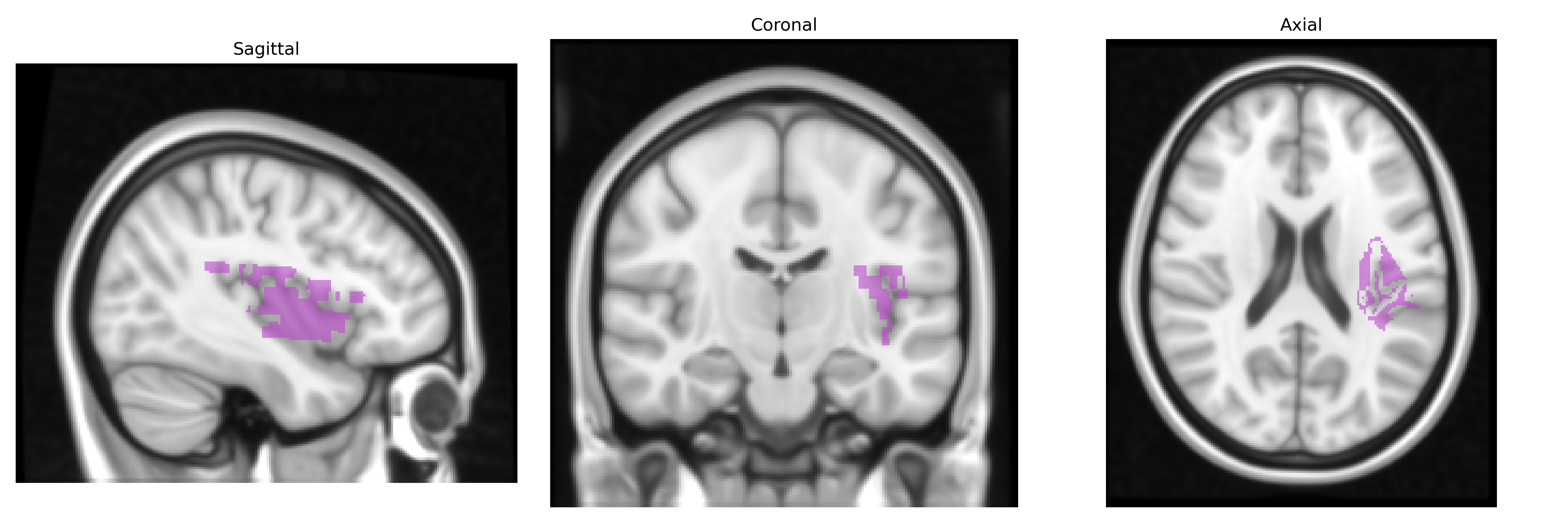
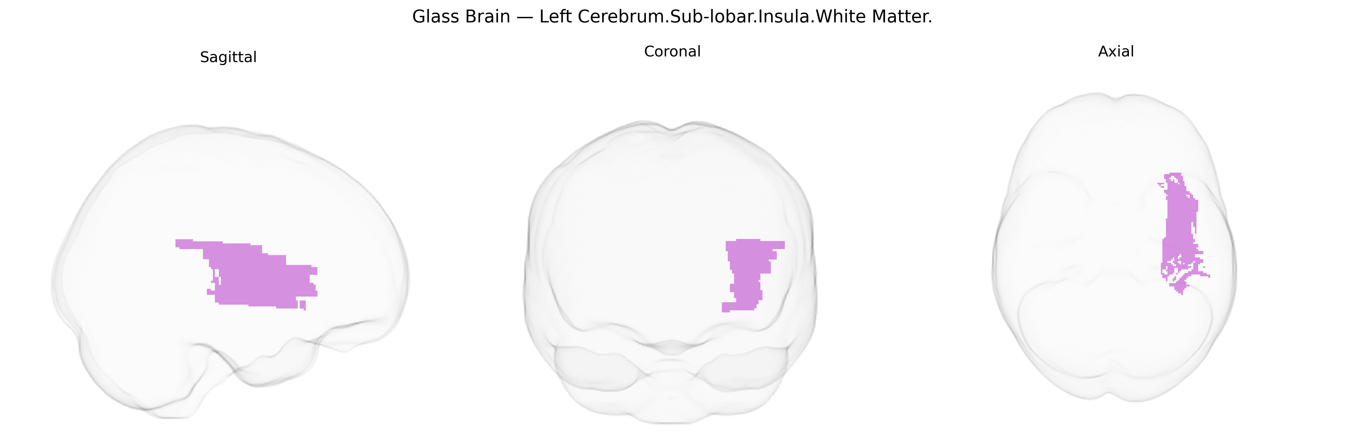
The bilateral Left Cerebrum.Sub-lobar.Insula.White Matter corresponds to the deep myelinated fiber tracts underlying the insular cortex within the sublobar region of the left cerebral hemisphere. These white matter pathways interconnect the insular gray matter with diverse cortical and subcortical targets, including frontal, temporal, and parietal association areas, limbic structures, and thalamic nuclei, thereby supporting integration of interoceptive, emotional, autonomic, and sensory information. Positioned deep within the lateral sulcus and covered by the opercular portions of the frontal, parietal, and temporal lobes, this white matter includes components of short U-fibers, association fibers, and projections that participate in salience, viscerosensory, and pain-processing networks. There is no direct link; a related structure is the Insular cortex.
The bilateral left insular white matter, as defined in the Talairach 1 mm atlas, participates in fronto-insular and temporo-insular connectivity that has been repeatedly implicated in imaging genetics and GWAS of brain structure and neuropsychiatric disease, although most studies report effects at the level of insular cortical thickness, surface area, or regional white-matter microstructure rather than this specific sub-lobar parcel. Large-scale neuroimaging GWAS (for example, ENIGMA and UK Biobank–based studies) have identified polygenic influences on insular volume and diffusion metrics, including common variants in or near genes involved in axon guidance, myelination, and synaptic function (e.g., NRG1/NRG3, CNTN4/6, NCAM1, and several cell-adhesion and oligodendrocyte-related loci), with many signals overlapping those that influence global white-matter integrity. Insula-related white-matter measures show heritability and polygenic overlap with schizophrenia, bipolar disorder, major depression, and autism spectrum disorder, consistent with case–control findings of altered insular connectivity in these conditions, and with risk loci in pathways such as glutamatergic signaling, calcium-channel genes (e.g., CACNA1C), and synaptic scaffolding (e.g., DLG2). Additionally, variants associated with traits such as risk-taking, substance use, pain sensitivity, interoceptive awareness, and cardiovascular regulation show correlated effects on insula structure or connectivity, aligning with the insula’s role in salience processing and autonomic control. Overall, genetic associations with this region are polygenic and pleiotropic, with risk and trait loci influencing insular white-matter characteristics as part of broader networks rather than in isolation, and with no single locus uniquely or specifically assigned to the “Left Cerebrum.Sub-lobar.Insula.White Matter” Talairach label.
Overview generated by GPT-4o (2026).
Region ID: 439
Hemisphere: bilateral
Atlas: Talairach labels 1mm
Full Quality Version: Download MP4
Full Quality Version: Download MP4
Wali Sidiqyar*, Gaurav Rudravaram*, Elyssa M. McMaster, Trent M. Schwartz, Adam M. Saunders, Kurt G. Schilling, Bennett A. Landman "Introducing SPINS: A Shared Public Visualization Library of Neuroanatomical Structures." Medical Imaging with Deep Learning- short paper
This resource is licensed under CC0 1.0 Universal (Public Domain).